 Movie
Movie Controller

[English] 日本語
 Yorodumi
- PDB-7oju: Chaetomium thermophilum Naa50 GNAT-domain in complex with bisubst... -
+ Open data

- Basic information
| Entry | Database: PDB / ID: 7oju | |||||||||
|---|---|---|---|---|---|---|---|---|---|---|
| Title | Chaetomium thermophilum Naa50 GNAT-domain in complex with bisubstrate analogue CoA-Ac-MVNAL | |||||||||
 Components Components |
| |||||||||
 Keywords Keywords |  TRANSFERASE / N-alpha-acetyltransferase / GNAT-fold / Naa50 / Chaetomium thermophilum TRANSFERASE / N-alpha-acetyltransferase / GNAT-fold / Naa50 / Chaetomium thermophilum | |||||||||
| Function / homology | N-acetyltransferase activity / Gcn5-related N-acetyltransferase (GNAT) domain profile. / GNAT domain / Acyl-CoA N-acyltransferase / CARBOXYMETHYL COENZYME *A / N-acetyltransferase-like protein Function and homology information Function and homology information | |||||||||
| Biological species |  Chaetomium thermophilum (fungus) Chaetomium thermophilum (fungus)synthetic construct (others) | |||||||||
| Method | X-RAY DIFFRACTION / SYNCHROTRON / MOLECULAR REPLACEMENT / Resolution: 1.1 Å | |||||||||
 Authors Authors | Weidenhausen, J. / Kopp, J. / Sinning, I. | |||||||||
| Funding support |  Germany, 2items Germany, 2items
| |||||||||
 Citation Citation | Journal: Int J Mol Sci / Year: 2022 Title: Extended N-Terminal Acetyltransferase Naa50 in Filamentous Fungi Adds to Naa50 Diversity. Authors: Weidenhausen, J. / Kopp, J. / Ruger-Herreros, C. / Stein, F. / Haberkant, P. / Lapouge, K. / Sinning, I. | |||||||||
| History |
|
- Structure visualization
| Structure viewer | Molecule: MolmilJmol/JSmol |
|---|
- Downloads & links
-Download
| PDBx/mmCIF format | 7oju.cif.gz | 151.3 KB | Display | PDBx/mmCIF format |
|---|---|---|---|---|
| PDB format | pdb7oju.ent.gz | 120.8 KB | Display | PDB format |
| PDBx/mmJSON format | 7oju.json.gz | Tree view | PDBx/mmJSON format | |
| Others |  Other downloads Other downloads |
-Validation report
| Arichive directory | https://data.pdbj.org/pub/pdb/validation_reports/oj/7ojuftp://data.pdbj.org/pub/pdb/validation_reports/oj/7oju | HTTPS FTP |
|---|
-Related structure data
| Related structure data | 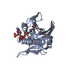 2ob0S  7ojv S: Starting model for refinement |
|---|---|
| Similar structure data |
-Links
 PDBj
PDBj

- Assembly
| Deposited unit | 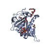
| ||||||||
|---|---|---|---|---|---|---|---|---|---|
| 1 |
| ||||||||
| Unit cell |
|
-Components
-Protein / Protein/peptide , 2 types, 2 molecules AH
| #1: Protein | Mass: 23732.049 Da / Num. of mol.: 1 / Mutation: N-terminal 81 aa & C-terminal 156 aa deletions Source method: isolated from a genetically manipulated source Details: Numbering is wrong: The protein was cloned with N- (81 aa) and C-terminal (156 aa) deletions. Therefore, the first leucine in the cloned sequence corresponds to residue number 82 (and not 3). ...Details: Numbering is wrong: The protein was cloned with N- (81 aa) and C-terminal (156 aa) deletions. Therefore, the first leucine in the cloned sequence corresponds to residue number 82 (and not 3). The N-terminal MG are due to cloning. Hexahistidine at the C-terminus is a His-tag. Source: (gene. exp.) Chaetomium thermophilum (strain DSM 1495 / CBS 144.50 / IMI 039719) (fungus)Strain: DSM 1495 / CBS 144.50 / IMI 039719 / Gene: CTHT_0014970 / Plasmid: pET24d / Production host:  Escherichia coli BL21(DE3) (bacteria) / Variant (production host): Rosetta2 / References: UniProt: G0S1V5 Escherichia coli BL21(DE3) (bacteria) / Variant (production host): Rosetta2 / References: UniProt: G0S1V5 |
|---|---|
| #2: Protein/peptide | Mass: 546.680 Da / Num. of mol.: 1 / Source method: obtained synthetically / Source: (synth.) synthetic construct (others) |
-Non-polymers , 4 types, 230 molecules 


| #3: Chemical | ChemComp-GOL / Glycerol Mass: 92.094 Da / Num. of mol.: 5 / Source method: obtained synthetically / Formula: C3H8O3 Mass: 92.094 Da / Num. of mol.: 5 / Source method: obtained synthetically / Formula: C3H8O3#4: Chemical | ChemComp-P6G / | Polyethylene glycol Mass: 282.331 Da / Num. of mol.: 1 / Source method: obtained synthetically / Formula: C12H26O7 / Comment: precipitant*YM Mass: 282.331 Da / Num. of mol.: 1 / Source method: obtained synthetically / Formula: C12H26O7 / Comment: precipitant*YM#5: Chemical | ChemComp-CMC / |  Mass: 825.570 Da / Num. of mol.: 1 / Source method: obtained synthetically / Formula: C23H38N7O18P3S / Feature type: SUBJECT OF INVESTIGATION Mass: 825.570 Da / Num. of mol.: 1 / Source method: obtained synthetically / Formula: C23H38N7O18P3S / Feature type: SUBJECT OF INVESTIGATION#6: Water | ChemComp-HOH / | WaterMass: 18.015 Da / Num. of mol.: 223 / Source method: isolated from a natural source / Formula: H2O |
|---|
-Details
| Has ligand of interest | Y |
|---|
-Experimental details
-Experiment
| Experiment | Method: X-RAY DIFFRACTION / Number of used crystals: 1 |
|---|
- Sample preparation
| Crystal | Density Matthews: 2.38 Å3/Da / Density % sol: 48.29 % |
|---|---|
| Crystal grow | Temperature: 291.15 K / Method: vapor diffusion, sitting drop / pH: 6 Details: 62 mg mL-1 protein mixed 1:2 with CoA-Ac-Lys; drops (600 nL): 1:1 mix of (20 mM HEPES pH 7.5, 500 mM NaCl) and (0.1 MES pH 6.0, 8 % (w/v) PEG3000, 37 % (v/v) PEG400). Crystals appeared after ...Details: 62 mg mL-1 protein mixed 1:2 with CoA-Ac-Lys; drops (600 nL): 1:1 mix of (20 mM HEPES pH 7.5, 500 mM NaCl) and (0.1 MES pH 6.0, 8 % (w/v) PEG3000, 37 % (v/v) PEG400). Crystals appeared after 1-2 days; 20 % (v/v) glycerol as cryo protectant |
-Data collection
| Diffraction | Mean temperature: 100 K / Serial crystal experiment: N |
|---|---|
| Diffraction source | Source: SYNCHROTRON / Site: PETRA III, EMBL c/o DESY / Beamline: P13 (MX1) / Wavelength: 0.976246 Å |
| Detector | Type: DECTRIS PILATUS 6M / Detector: PIXEL / Date: Mar 9, 2019 |
| Radiation | Monochromator: Si(111) / Protocol: SINGLE WAVELENGTH / Monochromatic (M) / Laue (L): M / Scattering type: x-ray |
| Radiation wavelength | Wavelength: 0.976246 Å / Relative weight: 1 |
| Reflection | Resolution: 1.1→41.67 Å / Num. obs: 92854 / % possible obs: 99.9 % / Redundancy: 12.7 % / CC1/2: 1 / Net I/σ(I): 18.01 |
| Reflection shell | Resolution: 1.1→1.14 Å / Redundancy: 12.5 % / Mean I/σ(I) obs: 1.45 / Num. unique obs: 9100 / CC1/2: 0.631 / % possible all: 99.7 |
- Processing
| Software |
| |||||||||||||||||||||||||||||||||||||||||||||||||||||||||||||||||||||||||||||||||||||||||||||||||||||||||||||||||||||||||||||||||||||||||||||||||||||||||||||||||||||||||||||||||||||||||||||||||||||||||||||||||||||||||
|---|---|---|---|---|---|---|---|---|---|---|---|---|---|---|---|---|---|---|---|---|---|---|---|---|---|---|---|---|---|---|---|---|---|---|---|---|---|---|---|---|---|---|---|---|---|---|---|---|---|---|---|---|---|---|---|---|---|---|---|---|---|---|---|---|---|---|---|---|---|---|---|---|---|---|---|---|---|---|---|---|---|---|---|---|---|---|---|---|---|---|---|---|---|---|---|---|---|---|---|---|---|---|---|---|---|---|---|---|---|---|---|---|---|---|---|---|---|---|---|---|---|---|---|---|---|---|---|---|---|---|---|---|---|---|---|---|---|---|---|---|---|---|---|---|---|---|---|---|---|---|---|---|---|---|---|---|---|---|---|---|---|---|---|---|---|---|---|---|---|---|---|---|---|---|---|---|---|---|---|---|---|---|---|---|---|---|---|---|---|---|---|---|---|---|---|---|---|---|---|---|---|---|---|---|---|---|---|---|---|---|---|---|---|---|---|---|---|---|
| Refinement | Method to determine structure: MOLECULAR REPLACEMENT Starting model: 2OB0 Resolution: 1.1→41.67 Å / SU ML: 0.1 / Cross valid method: THROUGHOUT / σ(F): 1.34 / Phase error: 14.61 / Stereochemistry target values: ML
| |||||||||||||||||||||||||||||||||||||||||||||||||||||||||||||||||||||||||||||||||||||||||||||||||||||||||||||||||||||||||||||||||||||||||||||||||||||||||||||||||||||||||||||||||||||||||||||||||||||||||||||||||||||||||
| Solvent computation | Shrinkage radii: 0.9 Å / VDW probe radii: 1.11 Å / Solvent model: FLAT BULK SOLVENT MODEL | |||||||||||||||||||||||||||||||||||||||||||||||||||||||||||||||||||||||||||||||||||||||||||||||||||||||||||||||||||||||||||||||||||||||||||||||||||||||||||||||||||||||||||||||||||||||||||||||||||||||||||||||||||||||||
| Displacement parameters | Biso max: 97.96 Å2 / Biso mean: 21.93 Å2 / Biso min: 7.94 Å2 | |||||||||||||||||||||||||||||||||||||||||||||||||||||||||||||||||||||||||||||||||||||||||||||||||||||||||||||||||||||||||||||||||||||||||||||||||||||||||||||||||||||||||||||||||||||||||||||||||||||||||||||||||||||||||
| Refinement step | Cycle: final / Resolution: 1.1→41.67 Å
| |||||||||||||||||||||||||||||||||||||||||||||||||||||||||||||||||||||||||||||||||||||||||||||||||||||||||||||||||||||||||||||||||||||||||||||||||||||||||||||||||||||||||||||||||||||||||||||||||||||||||||||||||||||||||
| LS refinement shell | Refine-ID: X-RAY DIFFRACTION / Rfactor Rfree error: 0 / Total num. of bins used: 30
|
