 Movie
Movie Controller


+ Open data

- Basic information
| Entry | Database: PDB / ID: 7og5 | ||||||
|---|---|---|---|---|---|---|---|
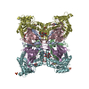
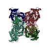
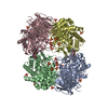
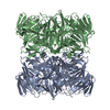
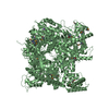
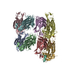
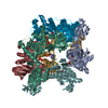
| Title | RNA-free Ribonuclease P from Halorhodospira halophila | ||||||
 Components Components | RNA-free ribonuclease P | ||||||
 Keywords Keywords |  HYDROLASE / RNAseP / metallonuclease / HARP HYDROLASE / RNAseP / metallonuclease / HARP | ||||||
| Function / homology | RNA-free ribonuclease P / PINc domain ribonuclease / ribonuclease P / ribonuclease P activity / tRNA 5'-leader removal / PIN-like domain superfamily / RNA-free ribonuclease P Function and homology information Function and homology information | ||||||
| Biological species |  Halorhodospira halophila (bacteria) Halorhodospira halophila (bacteria) | ||||||
| Method | ELECTRON MICROSCOPY / single particle reconstruction / cryo EM / Resolution: 3.37 Å | ||||||
 Authors Authors | Altegoer, F. / Bange, G. | ||||||
 Citation Citation | Journal: Elife / Year: 2021 Title: Structure and mechanistic features of the prokaryotic minimal RNase P. Authors: Rebecca Feyh / Nadine B Waeber / Simone Prinz / Pietro Ivan Giammarinaro / Gert Bange / Georg Hochberg / Roland K Hartmann / Florian Altegoer /  Abstract: Endonucleolytic removal of 5'-leader sequences from tRNA precursor transcripts (pre-tRNAs) by ribonuclease P (RNase P) is essential for protein synthesis. Beyond RNA-based RNase P enzymes, protein- ...Endonucleolytic removal of 5'-leader sequences from tRNA precursor transcripts (pre-tRNAs) by ribonuclease P (RNase P) is essential for protein synthesis. Beyond RNA-based RNase P enzymes, protein-only versions of the enzyme exert this function in various eukarya (there termed PRORPs) and in some bacteria ( and close relatives); both enzyme types belong to distinct subgroups of the PIN domain metallonuclease superfamily. Homologs of RNase P (HARPs) are also expressed in some other bacteria and many archaea, where they coexist with RNA-based RNase P and do not represent the main RNase P activity. Here, we solved the structure of the bacterial HARP from by cryo-electron microscopy, revealing a novel screw-like dodecameric assembly. Biochemical experiments demonstrate that oligomerization is required for RNase P activity of HARPs. We propose that the tRNA substrate binds to an extended spike-helix (SH) domain that protrudes from the screw-like assembly to position the 5'-end in close proximity to the active site of the neighboring dimer. The structure suggests that eukaryotic PRORPs and prokaryotic HARPs recognize the same structural elements of pre-tRNAs (tRNA elbow region and cleavage site). Our analysis thus delivers the structural and mechanistic basis for pre-tRNA processing by the prokaryotic HARP system. | ||||||
| History |
|
- Structure visualization
| Movie |
Movie viewer |
|---|---|
| Structure viewer | Molecule: MolmilJmol/JSmol |
- Downloads & links
-Download
| PDBx/mmCIF format | 7og5.cif.gz | 347.2 KB | Display | PDBx/mmCIF format |
|---|---|---|---|---|
| PDB format | pdb7og5.ent.gz | 286 KB | Display | PDB format |
| PDBx/mmJSON format | 7og5.json.gz | Tree view | PDBx/mmJSON format | |
| Others |  Other downloads Other downloads |
-Validation report
| Arichive directory | https://data.pdbj.org/pub/pdb/validation_reports/og/7og5ftp://data.pdbj.org/pub/pdb/validation_reports/og/7og5 | HTTPS FTP |
|---|
-Related structure data
| Related structure data | 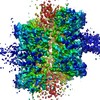 12878MC M: map data used to model this data C: citing same article ( |
|---|---|
| Similar structure data |

-Links
 PDBj
PDBj


- Assembly
| Deposited unit | 
|
|---|---|
| 1 |
|
-Components
| #1: Protein | Mass: 24051.338 Da / Num. of mol.: 12 Source method: isolated from a genetically manipulated source Source: (gene. exp.) Halorhodospira halophila (strain DSM 244 / SL1) (bacteria)Strain: DSM 244 / SL1 / Gene: Hhal_2243 / Production host: Escherichia coli BL21(DE3) (bacteria) / References: UniProt: A1WZ95, ribonuclease P |
|---|
-Experimental details
-Experiment
| Experiment | Method: ELECTRON MICROSCOPY |
|---|---|
| EM experiment | Aggregation state: PARTICLE / 3D reconstruction method: single particle reconstruction |
- Sample preparation
| Component | Name: Dodecameric assembly of minimal RNAseP system from Halorhodospira halophila Type: COMPLEX / Entity ID: all / Source: RECOMBINANT | |||||||||||||||
|---|---|---|---|---|---|---|---|---|---|---|---|---|---|---|---|---|
| Molecular weight | Value: 0.39 MDa / Experimental value: YES | |||||||||||||||
| Source (natural) | Organism: Halorhodospira halophila SL1 (bacteria) | |||||||||||||||
| Source (recombinant) | Organism: Escherichia coli BL21(DE3) (bacteria) | |||||||||||||||
| Buffer solution | pH: 8 Details: Solutions were prepared freshly and filtered through a 0.2 um filter | |||||||||||||||
| Buffer component |
| |||||||||||||||
| Specimen | Conc.: 8 mg/ml / Embedding applied: NO / Shadowing applied: NO / Staining applied: NO / Vitrification applied: YES / Details: The sample was monodisperse | |||||||||||||||
| Specimen support | Grid material: COPPER / Grid mesh size: 300 divisions/in. / Grid type: C-flat-1.2/1.3 | |||||||||||||||
| Vitrification | Instrument: FEI VITROBOT MARK IV / Cryogen name: ETHANE / Humidity: 100 % / Chamber temperature: 283 K / Details: Blot for 11s with blot force -1 before plunging |
- Electron microscopy imaging
| Experimental equipment |  Model: Titan Krios / Image courtesy: FEI Company |
|---|---|
| Microscopy | Model: FEI TITAN KRIOS |
| Electron gun | Electron source: FIELD EMISSION GUN / Accelerating voltage: 300 kV / Illumination mode: FLOOD BEAM |
| Electron lens | Mode: BRIGHT FIELDBright-field microscopy |
| Specimen holder | Cryogen: NITROGEN / Specimen holder model: FEI TITAN KRIOS AUTOGRID HOLDER |
| Image recording | Electron dose: 40 e/Å2 / Film or detector model: GATAN K3 (6k x 4k) / Num. of grids imaged: 1 / Num. of real images: 8393 |
- Processing
| Software |
| ||||||||||||||||||||||||
|---|---|---|---|---|---|---|---|---|---|---|---|---|---|---|---|---|---|---|---|---|---|---|---|---|---|
| EM software |
| ||||||||||||||||||||||||
| CTF correction | Type: PHASE FLIPPING AND AMPLITUDE CORRECTION | ||||||||||||||||||||||||
| 3D reconstruction | Resolution: 3.37 Å / Resolution method: FSC 0.143 CUT-OFF / Num. of particles: 1736597 / Symmetry type: POINT | ||||||||||||||||||||||||
| Atomic model building | B value: 181 / Protocol: AB INITIO MODEL / Space: REAL | ||||||||||||||||||||||||
| Refinement | Cross valid method: NONE Stereochemistry target values: GeoStd + Monomer Library + CDL v1.2 | ||||||||||||||||||||||||
| Displacement parameters | Biso mean: 106.7 Å2 | ||||||||||||||||||||||||
| Refine LS restraints |
|
