 Movie
Movie Controller

[English] 日本語
 Yorodumi
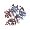
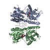
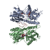
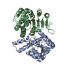
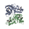
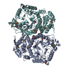
- PDB-7odc: CRYSTAL STRUCTURE ORNITHINE DECARBOXYLASE FROM MOUSE, TRUNCATED 3... -
+ Open data

- Basic information
| Entry | Database: PDB / ID: 7odc | ||||||
|---|---|---|---|---|---|---|---|
| Title | CRYSTAL STRUCTURE ORNITHINE DECARBOXYLASE FROM MOUSE, TRUNCATED 37 RESIDUES FROM THE C-TERMINUS, TO 1.6 ANGSTROM RESOLUTION | ||||||
 Components Components | PROTEIN (ORNITHINE DECARBOXYLASE) | ||||||
 Keywords Keywords |  LYASE / PYRIDOXAL-5'-PHOSPHATE / PLP / GROUP IV DECARBOXYLASE / POLYAMINES / PARASITICAL / CHEMOTHERAPY TARGET / ORNITHINE / PUTRESCINE / A/B-BARREL / OBLIGATE LYASE / PYRIDOXAL-5'-PHOSPHATE / PLP / GROUP IV DECARBOXYLASE / POLYAMINES / PARASITICAL / CHEMOTHERAPY TARGET / ORNITHINE / PUTRESCINE / A/B-BARREL / OBLIGATE | ||||||
| Function / homology |  Function and homology information Function and homology informationMetabolism of polyamines / putrescine biosynthetic process / putrescine biosynthetic process from ornithine / Regulation of ornithine decarboxylase (ODC) / ornithine decarboxylase / ornithine decarboxylase activity / polyamine metabolic process / regulation of protein catabolic process / kidney development / positive regulation of cell population proliferation ...Metabolism of polyamines / putrescine biosynthetic process / putrescine biosynthetic process from ornithine / Regulation of ornithine decarboxylase (ODC) / ornithine decarboxylase / ornithine decarboxylase activity / polyamine metabolic process / regulation of protein catabolic process / kidney development / positive regulation of cell population proliferation / perinuclear region of cytoplasm / protein homodimerization activity / cytosol / cytoplasmSimilarity search - Function | ||||||
| Biological species |  Mus musculus (house mouse) Mus musculus (house mouse) | ||||||
| Method | X-RAY DIFFRACTION / SYNCHROTRON / MIR / Resolution: 1.6 Å | ||||||
 Authors Authors | Kern, A.D. / Oliveira, M.A. / Coffino, P. / Hackert, M.L. | ||||||
 Citation Citation | Journal: Structure Fold.Des. / Year: 1999 Title: Structure of mammalian ornithine decarboxylase at 1.6 A resolution: stereochemical implications of PLP-dependent amino acid decarboxylases. Authors: Kern, A.D. / Oliveira, M.A. / Coffino, P. / Hackert, M.L. | ||||||
| History |
|
- Structure visualization
| Structure viewer | Molecule: MolmilJmol/JSmol |
|---|
- Downloads & links
-Download
| PDBx/mmCIF format | 7odc.cif.gz | 97.9 KB | Display | PDBx/mmCIF format |
|---|---|---|---|---|
| PDB format | pdb7odc.ent.gz | 73.9 KB | Display | PDB format |
| PDBx/mmJSON format | 7odc.json.gz | Tree view | PDBx/mmJSON format | |
| Others |  Other downloads Other downloads |
-Validation report
| Arichive directory | https://data.pdbj.org/pub/pdb/validation_reports/od/7odcftp://data.pdbj.org/pub/pdb/validation_reports/od/7odc | HTTPS FTP |
|---|
-Related structure data
| Similar structure data |
|---|


-Links
 PDBj
PDBj
- Assembly
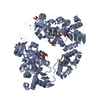
| Deposited unit | 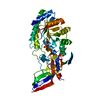
| ||||||||
|---|---|---|---|---|---|---|---|---|---|
| 1 | 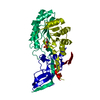
| ||||||||
| Unit cell |
|
-Components
| #1: Protein | Mass: 47275.801 Da / Num. of mol.: 1 / Mutation: TRUNCATED AFTER RESIDUE 424 Source method: isolated from a genetically manipulated source Details: SCHIFF-BASE LINKAGE BETWEEN PLP AND K69 / Source: (gene. exp.) Mus musculus (house mouse) / Tissue: LYMPHOMADescription: THE GENE WAS CLONED FROM A BALB-C MOUSE LYMPHOMA CELL LINE IN WHICH THE ODC GENE WAS AMPLIFIED BY SELECTION WITH DFMO. THE CELL LINE OF ORIGIN WAS S49.1, OBTAINED FROM THE SALK INSTITUTE ...Description: THE GENE WAS CLONED FROM A BALB-C MOUSE LYMPHOMA CELL LINE IN WHICH THE ODC GENE WAS AMPLIFIED BY SELECTION WITH DFMO. THE CELL LINE OF ORIGIN WAS S49.1, OBTAINED FROM THE SALK INSTITUTE CELL CULTURE CENTER IN 1974. THE ODC- AMPLIFIED LYMPHOMA CELL LINE OBTAINED AS A RESULT OF SELECTION WITH DFMO WAS D4. THE GENE HAS BEEN CLONED FROM MANY MOUSE STRAINS SINCE, AND THE ORF IS INVARIANT AMONG THESE, SO FAR AS WE KNOW. THE EXPRESSION OF THE PROTEIN WAS IN E.COLI. Cell line: S49.1 / Production host:  Escherichia coli (E. coli) / References: UniProt: P00860, ornithine decarboxylase Escherichia coli (E. coli) / References: UniProt: P00860, ornithine decarboxylase |
|---|---|
| #2: Chemical | ChemComp-PLP / Pyridoxal phosphate  Mass: 247.142 Da / Num. of mol.: 1 / Source method: obtained synthetically / Formula: C8H10NO6P Mass: 247.142 Da / Num. of mol.: 1 / Source method: obtained synthetically / Formula: C8H10NO6P |
| #3: Water | ChemComp-HOH / Water Mass: 18.015 Da / Num. of mol.: 421 / Source method: isolated from a natural source / Formula: H2O Mass: 18.015 Da / Num. of mol.: 421 / Source method: isolated from a natural source / Formula: H2O |
| Nonpolymer details | SCHIFF-BASE LINK BETWEEN LYS_69_NZ AND PLP_69_C4A |
| Sequence details | THE ENZYME WHOSE STRUCTURE WAS SOLVED IS A TRUNCATED ENZYME AND DOES NOT CONTAIN RESIDUES 425 ...THE ENZYME WHOSE STRUCTURE WAS SOLVED IS A TRUNCATED ENZYME AND DOES NOT CONTAIN RESIDUES 425 THROUGH 461. RESIDUES WHICH ARE PRESENT IN THIS ENZYME, BUT WHICH HAD NO CORRESPOND |
-Experimental details
-Experiment
| Experiment | Method: X-RAY DIFFRACTION / Number of used crystals: 1 |
|---|
- Sample preparation
| Crystal | Density Matthews: 2 Å3/Da / Density % sol: 37 % Description: DATA WERE COLLECTED USING THE OSCILLATION METHOD | ||||||||||||||||||||||||||||||
|---|---|---|---|---|---|---|---|---|---|---|---|---|---|---|---|---|---|---|---|---|---|---|---|---|---|---|---|---|---|---|---|
| Crystal grow | pH: 6.5 / Details: pH 6.5 | ||||||||||||||||||||||||||||||
| Crystal | *PLUS | ||||||||||||||||||||||||||||||
| Crystal grow | *PLUS Method: vapor diffusion / Details: combination of vapor diffusion and gel | ||||||||||||||||||||||||||||||
| Components of the solutions | *PLUS
|
-Data collection
| Diffraction | Mean temperature: 98 K |
|---|---|
| Diffraction source | Source: SYNCHROTRON / Site: CHESS  / Beamline: F1 / Wavelength: 0.908 / Beamline: F1 / Wavelength: 0.908 |
| Detector | Detector: CCD / Date: Sep 1, 1996 |
| Radiation | Protocol: SINGLE WAVELENGTH / Monochromatic (M) / Laue (L): M / Scattering type: x-ray |
| Radiation wavelength | Wavelength: 0.908 Å / Relative weight: 1 |
| Reflection | Resolution: 1.6→27.92 Å / Num. obs: 49125 / % possible obs: 91.3 % / Redundancy: 5 % / Rmerge(I) obs: 0.081 |
| Reflection shell | Resolution: 1.6→1.66 Å / Redundancy: 1.7 % / Rmerge(I) obs: 0.188 / Mean I/σ(I) obs: 3.5 / % possible all: 75.4 |
| Reflection shell | *PLUS % possible obs: 75.4 % |
- Processing
| Software |
| |||||||||||||||||||||
|---|---|---|---|---|---|---|---|---|---|---|---|---|---|---|---|---|---|---|---|---|---|---|
| Refinement | Method to determine structure: MIR / Resolution: 1.6→27.9 Å / Cross valid method: THROUGHOUT / σ(F): 0 Details: THE PROGRAM USED FOR REFINEMENT WAS CCP PROGRAM SUITE: REFMAC_3.3 VERSION 3.4
| |||||||||||||||||||||
| Refinement step | Cycle: LAST / Resolution: 1.6→27.9 Å
| |||||||||||||||||||||
| Software | *PLUS Name: REFMAC / Classification: refinement | |||||||||||||||||||||
| Refinement | *PLUS Highest resolution: 1.6 Å / Lowest resolution: 27.9 Å / σ(F): 0 / % reflection Rfree: 4 % / Rfactor obs: 0.2 | |||||||||||||||||||||
| Solvent computation | *PLUS | |||||||||||||||||||||
| Displacement parameters | *PLUS | |||||||||||||||||||||
| Refine LS restraints | *PLUS
|
