 Movie
Movie Controller

[English] 日本語
 Yorodumi
- PDB-7o5h: Ribosomal methyltransferase KsgA bound to small ribosomal subunit -
+ Open data

- Basic information
| Entry | Database: PDB / ID: 7o5h | ||||||
|---|---|---|---|---|---|---|---|
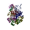
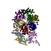
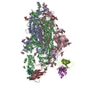
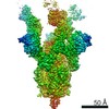
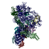
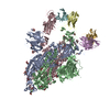
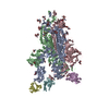
| Title | Ribosomal methyltransferase KsgA bound to small ribosomal subunit | ||||||
 Components Components |
| ||||||
 Keywords Keywords |  RIBOSOME / Methyltransferase KsgA / 30S RIBOSOME / Methyltransferase KsgA / 30S | ||||||
| Function / homology |  Function and homology information16S rRNA (adenine1518-N6/adenine1519-N6)-dimethyltransferase / 16S rRNA (adenine(1518)-N(6)/adenine(1519)-N(6))-dimethyltransferase activity / mRNA 5'-UTR binding / small ribosomal subunit rRNA binding / ribosomal small subunit assembly / cytosolic small ribosomal subunit / cytoplasmic translation / small ribosomal subunit / tRNA binding / rRNA binding ...16S rRNA (adenine1518-N6/adenine1519-N6)-dimethyltransferase / 16S rRNA (adenine(1518)-N(6)/adenine(1519)-N(6))-dimethyltransferase activity / mRNA 5'-UTR binding / small ribosomal subunit rRNA binding / ribosomal small subunit assembly / cytosolic small ribosomal subunit / cytoplasmic translation / small ribosomal subunit / tRNA binding / rRNA binding / ribosome / structural constituent of ribosome / ribonucleoprotein complex / translation / RNA binding / cytosol / cytoplasm Function and homology information16S rRNA (adenine1518-N6/adenine1519-N6)-dimethyltransferase / 16S rRNA (adenine(1518)-N(6)/adenine(1519)-N(6))-dimethyltransferase activity / mRNA 5'-UTR binding / small ribosomal subunit rRNA binding / ribosomal small subunit assembly / cytosolic small ribosomal subunit / cytoplasmic translation / small ribosomal subunit / tRNA binding / rRNA binding ...16S rRNA (adenine1518-N6/adenine1519-N6)-dimethyltransferase / 16S rRNA (adenine(1518)-N(6)/adenine(1519)-N(6))-dimethyltransferase activity / mRNA 5'-UTR binding / small ribosomal subunit rRNA binding / ribosomal small subunit assembly / cytosolic small ribosomal subunit / cytoplasmic translation / small ribosomal subunit / tRNA binding / rRNA binding / ribosome / structural constituent of ribosome / ribonucleoprotein complex / translation / RNA binding / cytosol / cytoplasmSimilarity search - Function | ||||||
| Biological species |  Escherichia coli (E. coli) Escherichia coli (E. coli) | ||||||
| Method | ELECTRON MICROSCOPY / single particle reconstruction / cryo EM / Resolution: 3.1 Å | ||||||
 Authors Authors | Stephan, N.C. / Ries, A.B. / Boehringer, D. / Ban, N. | ||||||
| Funding support |  Switzerland, 1items Switzerland, 1items
| ||||||
 Citation Citation | Journal: Nucleic Acids Res / Year: 2021 Title: Structural basis of successive adenosine modifications by the conserved ribosomal methyltransferase KsgA. Authors: Niklas C Stephan / Anne B Ries / Daniel Boehringer / Nenad Ban / Abstract: Biogenesis of ribosomal subunits involves enzymatic modifications of rRNA that fine-tune functionally important regions. The universally conserved prokaryotic dimethyltransferase KsgA sequentially ...Biogenesis of ribosomal subunits involves enzymatic modifications of rRNA that fine-tune functionally important regions. The universally conserved prokaryotic dimethyltransferase KsgA sequentially modifies two universally conserved adenosine residues in helix 45 of the small ribosomal subunit rRNA, which is in proximity of the decoding site. Here we present the cryo-EM structure of Escherichia coli KsgA bound to an E. coli 30S at a resolution of 3.1 Å. The high-resolution structure reveals how KsgA recognizes immature rRNA and binds helix 45 in a conformation where one of the substrate nucleotides is flipped-out into the active site. We suggest that successive processing of two adjacent nucleotides involves base-flipping of the rRNA, which allows modification of the second substrate nucleotide without dissociation of the enzyme. Since KsgA is homologous to the essential eukaryotic methyltransferase Dim1 involved in 40S maturation, these results have also implications for understanding eukaryotic ribosome maturation. | ||||||
| History |
|
- Structure visualization
| Movie |
Movie viewer |
|---|---|
| Structure viewer | Molecule: MolmilJmol/JSmol |
- Downloads & links
-Download
| PDBx/mmCIF format | 7o5h.cif.gz | 797.3 KB | Display | PDBx/mmCIF format |
|---|---|---|---|---|
| PDB format | pdb7o5h.ent.gz | 612.8 KB | Display | PDB format |
| PDBx/mmJSON format | 7o5h.json.gz | Tree view | PDBx/mmJSON format | |
| Others |  Other downloads Other downloads |
-Validation report
| Arichive directory | https://data.pdbj.org/pub/pdb/validation_reports/o5/7o5hftp://data.pdbj.org/pub/pdb/validation_reports/o5/7o5h | HTTPS FTP |
|---|
-Related structure data
| Related structure data | 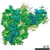 12736MC M: map data used to model this data C: citing same article ( |
|---|---|
| Similar structure data |
-Links
 PDBj
PDBj


- Assembly
| Deposited unit | 
|
|---|---|
| 1 |
|
-Components
-30S ribosomal protein ... , 13 types, 13 molecules BDEFHKLOPQRTU
| #3: Protein | Mass: 25072.867 Da / Num. of mol.: 1 / Source method: isolated from a natural source / Source: (natural) Escherichia coli (E. coli) / Strain: JW0050.3 / References: UniProt: A0A3K3SDJ5 |
|---|---|
| #4: Protein | Mass: 23383.002 Da / Num. of mol.: 1 / Source method: isolated from a natural source / Source: (natural) Escherichia coli (E. coli) / Strain: JW0050.3 / References: UniProt: V0YKB3 |
| #5: Protein | Mass: 16661.268 Da / Num. of mol.: 1 / Source method: isolated from a natural source / Source: (natural) Escherichia coli (E. coli) / Strain: JW0050.3 / References: UniProt: A1AGJ2 |
| #6: Protein | Ribosome Mass: 12326.251 Da / Num. of mol.: 1 / Source method: isolated from a natural source / Source: (natural) Escherichia coli (E. coli) / Strain: JW0050.3 / References: UniProt: P02358 |
| #7: Protein | Mass: 14015.361 Da / Num. of mol.: 1 / Source method: isolated from a natural source / Source: (natural) Escherichia coli (E. coli) / Strain: JW0050.3 / References: UniProt: H4JDM0 |
| #8: Protein | Mass: 12487.200 Da / Num. of mol.: 1 / Source method: isolated from a natural source / Source: (natural) Escherichia coli (E. coli) / Strain: JW0050.3 / References: UniProt: A0A5I5F5V9 |
| #9: Protein | Mass: 13636.961 Da / Num. of mol.: 1 / Source method: isolated from a natural source / Source: (natural) Escherichia coli (E. coli) / Strain: JW0050.3 / References: UniProt: I2UHF1 |
| #10: Protein | Mass: 10159.621 Da / Num. of mol.: 1 / Source method: isolated from a natural source / Source: (natural) Escherichia coli (E. coli) / Strain: JW0050.3 / References: UniProt: E9YUR8 |
| #11: Protein | Mass: 9207.572 Da / Num. of mol.: 1 / Source method: isolated from a natural source / Source: (natural) Escherichia coli (E. coli) / Strain: JW0050.3 / References: UniProt: C3SYP2 |
| #12: Protein | Mass: 9263.946 Da / Num. of mol.: 1 / Source method: isolated from a natural source / Source: (natural) Escherichia coli (E. coli) / Strain: JW0050.3 / References: UniProt: T6LV72 |
| #13: Protein | Mass: 6466.477 Da / Num. of mol.: 1 / Source method: isolated from a natural source / Source: (natural) Escherichia coli (E. coli) / Strain: JW0050.3 / References: UniProt: A7ZV73 |
| #14: Protein | Mass: 9577.268 Da / Num. of mol.: 1 / Source method: isolated from a natural source / Source: (natural) Escherichia coli (E. coli) / Strain: JW0050.3 / References: UniProt: I4T5W9 |
| #15: Protein | Mass: 6629.744 Da / Num. of mol.: 1 / Source method: isolated from a natural source / Source: (natural) Escherichia coli (E. coli) / Strain: JW0050.3 / References: UniProt: A0A5C9AJ78 |
-Protein / RNA chain / Non-polymers , 3 types, 169 molecules VA
| #16: Chemical | ChemComp-MG /  Mass: 24.305 Da / Num. of mol.: 167 / Source method: obtained synthetically / Formula: Mg Mass: 24.305 Da / Num. of mol.: 167 / Source method: obtained synthetically / Formula: Mg#1: Protein | | / 16S rRNA (adenine(1518)-N(6)/adenine(1519)-N(6))-dimethyltransferase / 16S rRNA dimethyladenosine ...16S rRNA (adenine(1518)-N(6)/adenine(1519)-N(6))-dimethyltransferase / 16S rRNA dimethyladenosine transferase / 16S rRNA dimethylase / S-adenosylmethionine-6-N' / N'-adenosyl(rRNA) dimethyltransferaseMass: 27988.273 Da / Num. of mol.: 1 Source method: isolated from a genetically manipulated source Source: (gene. exp.) Escherichia coli (strain K12) (bacteria)Strain: K12 / Gene: rsmA, ksgA, FAZ83_07175 / Production host: Escherichia coli BL21(DE3) (bacteria)References: UniProt: A0A4S5B3V1, 16S rRNA (adenine1518-N6/adenine1519-N6)-dimethyltransferase#2: RNA chain | | Mass: 312980.562 Da / Num. of mol.: 1 / Source method: isolated from a natural source / Source: (natural) Escherichia coli (E. coli) / Strain: JW0050.3 |
|---|
-Details
| Has ligand of interest | N |
|---|
-Experimental details
-Experiment
| Experiment | Method: ELECTRON MICROSCOPY |
|---|---|
| EM experiment | Aggregation state: PARTICLE / 3D reconstruction method: single particle reconstruction |
- Sample preparation
| Component | Name: KsgA bound to bacterial ribosomal small subunit 30S / Type: RIBOSOME / Entity ID: #1-#15 / Source: MULTIPLE SOURCES |
|---|---|
| Source (natural) | Organism: Escherichia coli (E. coli) |
| Buffer solution | pH: 7.6 |
| Specimen | Embedding applied: NO / Shadowing applied: NO / Staining applied: NO / Vitrification applied: YES |
| Vitrification | Cryogen name: ETHANE-PROPANE |
- Electron microscopy imaging
| Experimental equipment |  Model: Titan Krios / Image courtesy: FEI Company |
|---|---|
| Microscopy | Model: FEI TITAN KRIOS |
| Electron gun | Electron source: FIELD EMISSION GUN / Accelerating voltage: 300 kV / Illumination mode: OTHER |
| Electron lens | Mode: BRIGHT FIELDBright-field microscopy |
| Image recording | Electron dose: 35 e/Å2 / Film or detector model: FEI FALCON III (4k x 4k) |
- Processing
| Software |
| ||||||||||||||||||||||||
|---|---|---|---|---|---|---|---|---|---|---|---|---|---|---|---|---|---|---|---|---|---|---|---|---|---|
| CTF correction | Type: PHASE FLIPPING AND AMPLITUDE CORRECTION | ||||||||||||||||||||||||
| 3D reconstruction | Resolution: 3.1 Å / Resolution method: FSC 0.143 CUT-OFF / Num. of particles: 165073 / Symmetry type: POINT | ||||||||||||||||||||||||
| Refinement | Cross valid method: NONE Stereochemistry target values: GeoStd + Monomer Library + CDL v1.2 | ||||||||||||||||||||||||
| Displacement parameters | Biso mean: 68.69 Å2 | ||||||||||||||||||||||||
| Refine LS restraints |
|
