 Movie
Movie Controller

[English] 日本語
 Yorodumi
- PDB-7nzq: D-lyxose isomerase from the hyperthermophilic archaeon Thermofilu... -
+ Open data

- Basic information
| Entry | Database: PDB / ID: 7nzq | ||||||
|---|---|---|---|---|---|---|---|
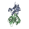
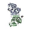
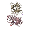
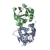
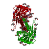
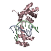
| Title | D-lyxose isomerase from the hyperthermophilic archaeon Thermofilum sp complexed with D-mannose | ||||||
 Components Components | D-lyxose/D-mannose family sugar isomerase | ||||||
 Keywords Keywords |  ISOMERASE / Complex / thermostability / biocatalysis ISOMERASE / Complex / thermostability / biocatalysis | ||||||
| Function / homology |  Function and homology information Function and homology information | ||||||
| Biological species |  Thermofilum sp. ex4484_79 (archaea) Thermofilum sp. ex4484_79 (archaea) | ||||||
| Method | X-RAY DIFFRACTION / SYNCHROTRON / MOLECULAR REPLACEMENT / Resolution: 1.57 Å | ||||||
 Authors Authors | De Rose, S.A. / Isupov, M.N. / Littlechild, J.A. / Schoenheit, P. | ||||||
| Funding support |  Germany, 1items Germany, 1items
| ||||||
 Citation Citation | Journal: Front Bioeng Biotechnol / Year: 2021 Title: Biochemical and Structural Characterisation of a Novel D-Lyxose Isomerase From the Hyperthermophilic Archaeon Thermofilum sp. Authors: De Rose, S.A. / Kuprat, T. / Isupov, M.N. / Reinhardt, A. / Schonheit, P. / Littlechild, J.A. | ||||||
| History |
|
- Structure visualization
| Structure viewer | Molecule: MolmilJmol/JSmol |
|---|
- Downloads & links
-Download
| PDBx/mmCIF format | 7nzq.cif.gz | 65 KB | Display | PDBx/mmCIF format |
|---|---|---|---|---|
| PDB format | pdb7nzq.ent.gz | Display | PDB format | |
| PDBx/mmJSON format | 7nzq.json.gz | Tree view | PDBx/mmJSON format | |
| Others |  Other downloads Other downloads |
-Validation report
| Arichive directory | https://data.pdbj.org/pub/pdb/validation_reports/nz/7nzqftp://data.pdbj.org/pub/pdb/validation_reports/nz/7nzq | HTTPS FTP |
|---|
-Related structure data
| Related structure data |  7nzoC  7nzpC  2y0oS S: Starting model for refinement C: citing same article ( |
|---|---|
| Similar structure data |


-Links
 PDBj
PDBj- Assembly
| Deposited unit | 
| ||||||||||||
|---|---|---|---|---|---|---|---|---|---|---|---|---|---|
| 1 | 
| ||||||||||||
| Unit cell |
| ||||||||||||
| Components on special symmetry positions |
|
-Components
-Protein / Sugars , 2 types, 2 molecules AAA
| #1: Protein | Mass: 23898.215 Da / Num. of mol.: 1 Source method: isolated from a genetically manipulated source Source: (gene. exp.) Thermofilum sp. ex4484_79 (archaea) / Gene: B6U94_07925 / Production host:  Escherichia coli (E. coli) / References: UniProt: A0A256XLS3 Escherichia coli (E. coli) / References: UniProt: A0A256XLS3 |
|---|---|
| #2: Sugar | ChemComp-BMA / Mannose Type: D-saccharide, beta linking / Mass: 180.156 Da / Num. of mol.: 1 / Source method: obtained synthetically / Formula: C6H12O6 / Feature type: SUBJECT OF INVESTIGATION Type: D-saccharide, beta linking / Mass: 180.156 Da / Num. of mol.: 1 / Source method: obtained synthetically / Formula: C6H12O6 / Feature type: SUBJECT OF INVESTIGATION |
-Non-polymers , 5 types, 229 molecules 


| #3: Chemical | Ethylene glycol Mass: 62.068 Da / Num. of mol.: 2 / Source method: obtained synthetically / Formula: C2H6O2 Mass: 62.068 Da / Num. of mol.: 2 / Source method: obtained synthetically / Formula: C2H6O2#4: Chemical | ChemComp-PEG / | Diethylene glycol Mass: 106.120 Da / Num. of mol.: 1 / Source method: obtained synthetically / Formula: C4H10O3 Mass: 106.120 Da / Num. of mol.: 1 / Source method: obtained synthetically / Formula: C4H10O3#5: Chemical | ChemComp-MN / |  Mass: 54.938 Da / Num. of mol.: 1 / Source method: obtained synthetically / Formula: Mn / Feature type: SUBJECT OF INVESTIGATION Mass: 54.938 Da / Num. of mol.: 1 / Source method: obtained synthetically / Formula: Mn / Feature type: SUBJECT OF INVESTIGATION#6: Chemical | ChemComp-PO4 / | Phosphate Mass: 94.971 Da / Num. of mol.: 1 / Source method: obtained synthetically / Formula: PO4 Mass: 94.971 Da / Num. of mol.: 1 / Source method: obtained synthetically / Formula: PO4#7: Water | ChemComp-HOH / | WaterMass: 18.015 Da / Num. of mol.: 224 / Source method: isolated from a natural source / Formula: H2O |
|---|
-Details
| Has ligand of interest | Y |
|---|
-Experimental details
-Experiment
| Experiment | Method: X-RAY DIFFRACTION / Number of used crystals: 1 |
|---|
- Sample preparation
| Crystal | Density Matthews: 2.46 Å3/Da / Density % sol: 49.91 % |
|---|---|
| Crystal grow | Temperature: 291 K / Method: microbatch / pH: 6 Details: 100 mM SPG ph 6.0, 25% w/v PEG 1500, 40 mM D-mannose |
-Data collection
| Diffraction | Mean temperature: 100 K / Serial crystal experiment: N |
|---|---|
| Diffraction source | Source: SYNCHROTRON / Site: Diamond  / Beamline: I04 / Wavelength: 0.9795 Å / Beamline: I04 / Wavelength: 0.9795 Å |
| Detector | Type: DECTRIS EIGER2 XE 16M / Detector: PIXEL / Date: Dec 15, 2019 |
| Radiation | Protocol: SINGLE WAVELENGTH / Monochromatic (M) / Laue (L): M / Scattering type: x-ray |
| Radiation wavelength | Wavelength: 0.9795 Å / Relative weight: 1 |
| Reflection | Resolution: 1.57→37.692 Å / Num. obs: 29074 / % possible obs: 99.9 % / Redundancy: 5.2 % / CC1/2: 0.997 / Net I/σ(I): 6.6 |
| Reflection shell | Resolution: 1.57→1.6 Å / Num. unique obs: 1425 / CC1/2: 0.277 |
- Processing
| Software |
| |||||||||||||||||||||||||||||||||||||||||||||||||||||||||||||||||||||||||||||||||||||||||||||||||||||||||||||||||||||||||||||||||||||||||||||||||||
|---|---|---|---|---|---|---|---|---|---|---|---|---|---|---|---|---|---|---|---|---|---|---|---|---|---|---|---|---|---|---|---|---|---|---|---|---|---|---|---|---|---|---|---|---|---|---|---|---|---|---|---|---|---|---|---|---|---|---|---|---|---|---|---|---|---|---|---|---|---|---|---|---|---|---|---|---|---|---|---|---|---|---|---|---|---|---|---|---|---|---|---|---|---|---|---|---|---|---|---|---|---|---|---|---|---|---|---|---|---|---|---|---|---|---|---|---|---|---|---|---|---|---|---|---|---|---|---|---|---|---|---|---|---|---|---|---|---|---|---|---|---|---|---|---|---|---|---|---|
| Refinement | Method to determine structure: MOLECULAR REPLACEMENT Starting model: 2y0o Resolution: 1.57→37.692 Å / Cor.coef. Fo:Fc: 0.973 / Cor.coef. Fo:Fc free: 0.951 / SU B: 2.575 / SU ML: 0.082 / Cross valid method: FREE R-VALUE / ESU R: 0.085 / ESU R Free: 0.09 / Details: Hydrogens have not been used
| |||||||||||||||||||||||||||||||||||||||||||||||||||||||||||||||||||||||||||||||||||||||||||||||||||||||||||||||||||||||||||||||||||||||||||||||||||
| Solvent computation | Ion probe radii: 0.8 Å / Shrinkage radii: 0.8 Å / VDW probe radii: 1.2 Å / Solvent model: BABINET MODEL PLUS MASK | |||||||||||||||||||||||||||||||||||||||||||||||||||||||||||||||||||||||||||||||||||||||||||||||||||||||||||||||||||||||||||||||||||||||||||||||||||
| Displacement parameters | Biso mean: 25.025 Å2
| |||||||||||||||||||||||||||||||||||||||||||||||||||||||||||||||||||||||||||||||||||||||||||||||||||||||||||||||||||||||||||||||||||||||||||||||||||
| Refinement step | Cycle: LAST / Resolution: 1.57→37.692 Å
| |||||||||||||||||||||||||||||||||||||||||||||||||||||||||||||||||||||||||||||||||||||||||||||||||||||||||||||||||||||||||||||||||||||||||||||||||||
| Refine LS restraints |
| |||||||||||||||||||||||||||||||||||||||||||||||||||||||||||||||||||||||||||||||||||||||||||||||||||||||||||||||||||||||||||||||||||||||||||||||||||
| LS refinement shell |
|
