 Movie
Movie Controller

[English] 日本語
 Yorodumi
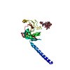
- PDB-7nvk: Crystal structure of UBA5 fragment fused to the N-terminus of UFC1 -
+ Open data

- Basic information
| Entry | Database: PDB / ID: 7nvk | ||||||
|---|---|---|---|---|---|---|---|
| Title | Crystal structure of UBA5 fragment fused to the N-terminus of UFC1 | ||||||
 Components Components | Ubiquitin-like modifier-activating enzyme 5,Ubiquitin-fold modifier-conjugating enzyme 1 | ||||||
 Keywords Keywords |  LIGASE / Ubiquitin like fold modifier enzyme 5 (UBA5) / E1 / Ubiquitin fold modifier 1 (UFM1) / Ubiquitin fold modifier conjugating enzyme 1 (UFC1) / UFMYLATION LIGASE / Ubiquitin like fold modifier enzyme 5 (UBA5) / E1 / Ubiquitin fold modifier 1 (UFM1) / Ubiquitin fold modifier conjugating enzyme 1 (UFC1) / UFMYLATION | ||||||
| Function / homology |  Function and homology information Function and homology informationUFM1 conjugating enzyme activity / UFM1 activating enzyme activity / UFM1 transferase activity / protein ufmylation / protein K69-linked ufmylation / megakaryocyte differentiation / regulation of intracellular estrogen receptor signaling pathway / reticulophagy / neuromuscular process / localization ...UFM1 conjugating enzyme activity / UFM1 activating enzyme activity / UFM1 transferase activity / protein ufmylation / protein K69-linked ufmylation / megakaryocyte differentiation / regulation of intracellular estrogen receptor signaling pathway / reticulophagy / neuromuscular process / localization / response to endoplasmic reticulum stress / erythrocyte differentiation / brain development / Antigen processing: Ubiquitination & Proteasome degradation / intracellular membrane-bounded organelle / endoplasmic reticulum membrane / Golgi apparatus / protein homodimerization activity / extracellular exosome / zinc ion binding / ATP binding / nucleus / cytosol / cytoplasmSimilarity search - Function | ||||||
| Biological species |  Homo sapiens (human) Homo sapiens (human) | ||||||
| Method | X-RAY DIFFRACTION / SYNCHROTRON / MOLECULAR REPLACEMENT / Resolution: 2.651 Å | ||||||
 Authors Authors | Manoj Kumar, P. / Padala, P. / Isupov, M.N. / Wiener, R. | ||||||
| Funding support |  Israel, 1items Israel, 1items
| ||||||
 Citation Citation | Journal: Nat Commun / Year: 2021 Title: Structural basis for UFM1 transfer from UBA5 to UFC1. Authors: Kumar, M. / Padala, P. / Fahoum, J. / Hassouna, F. / Tsaban, T. / Zoltsman, G. / Banerjee, S. / Cohen-Kfir, E. / Dessau, M. / Rosenzweig, R. / Isupov, M.N. / Schueler-Furman, O. / Wiener, R. | ||||||
| History |
|
- Structure visualization
| Structure viewer | Molecule: MolmilJmol/JSmol |
|---|
- Downloads & links
-Download
| PDBx/mmCIF format | 7nvk.cif.gz | 53.5 KB | Display | PDBx/mmCIF format |
|---|---|---|---|---|
| PDB format | pdb7nvk.ent.gz | Display | PDB format | |
| PDBx/mmJSON format | 7nvk.json.gz | Tree view | PDBx/mmJSON format | |
| Others |  Other downloads Other downloads |
-Validation report
| Arichive directory | https://data.pdbj.org/pub/pdb/validation_reports/nv/7nvkftp://data.pdbj.org/pub/pdb/validation_reports/nv/7nvk | HTTPS FTP |
|---|
-Related structure data
| Related structure data | 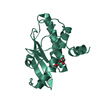 7nvjC 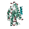 7nw1C  2z6oS S: Starting model for refinement C: citing same article ( |
|---|---|
| Similar structure data |


-Links
 PDBj
PDBj


- Assembly
| Deposited unit | 
| ||||||||
|---|---|---|---|---|---|---|---|---|---|
| 1 |
| ||||||||
| Unit cell |
|
-Components
| #1: Protein | Mass: 25917.533 Da / Num. of mol.: 1 Source method: isolated from a genetically manipulated source Source: (gene. exp.) Homo sapiens (human) / Gene: UBA5, UBE1DC1, UFC1, CGI-126, HSPC155 / Plasmid: pET32A / Production host:  Escherichia coli (E. coli) / References: UniProt: Q9GZZ9, UniProt: Q9Y3C8 Escherichia coli (E. coli) / References: UniProt: Q9GZZ9, UniProt: Q9Y3C8 |
|---|
-Experimental details
-Experiment
| Experiment | Method: X-RAY DIFFRACTION / Number of used crystals: 1 |
|---|
- Sample preparation
| Crystal | Density Matthews: 2.51 Å3/Da / Density % sol: 51 % |
|---|---|
| Crystal grow | Temperature: 293 K / Method: vapor diffusion, hanging drop / pH: 7.5 Details: 2% (v/v) Tacsimate pH 7.0, 20% PEG 3350, 0.1M HEPES pH 7.5, 6mM zinc sulfate |
-Data collection
| Diffraction | Mean temperature: 298 K / Serial crystal experiment: N |
|---|---|
| Diffraction source | Source: SYNCHROTRON / Site: BESSY  / Beamline: 14.1 / Wavelength: 0.9763 Å / Beamline: 14.1 / Wavelength: 0.9763 Å |
| Detector | Type: DECTRIS PILATUS3 6M / Detector: PIXEL / Date: Dec 20, 2019 |
| Radiation | Protocol: SINGLE WAVELENGTH / Monochromatic (M) / Laue (L): M / Scattering type: x-ray |
| Radiation wavelength | Wavelength: 0.9763 Å / Relative weight: 1 |
| Reflection | Resolution: 2.65→46.78 Å / Num. obs: 7314 / % possible obs: 100 % / Redundancy: 20.4 % / Biso Wilson estimate: 127.1 Å2 / CC1/2: 0.99 / Rrim(I) all: 0.112 / Net I/σ(I): 13.1 |
| Reflection shell | Resolution: 2.65→2.78 Å / Redundancy: 21.4 % / Num. unique obs: 953 / CC1/2: 0.292 / Rrim(I) all: 0.6 / % possible all: 100 |
- Processing
| Software |
| |||||||||||||||||||||||||||||||||||||||||||||||||||||||||||||||||||||||||||||||||||||||||||||||||||||||||||||||||||||||||||||||||||||||||||||||||||
|---|---|---|---|---|---|---|---|---|---|---|---|---|---|---|---|---|---|---|---|---|---|---|---|---|---|---|---|---|---|---|---|---|---|---|---|---|---|---|---|---|---|---|---|---|---|---|---|---|---|---|---|---|---|---|---|---|---|---|---|---|---|---|---|---|---|---|---|---|---|---|---|---|---|---|---|---|---|---|---|---|---|---|---|---|---|---|---|---|---|---|---|---|---|---|---|---|---|---|---|---|---|---|---|---|---|---|---|---|---|---|---|---|---|---|---|---|---|---|---|---|---|---|---|---|---|---|---|---|---|---|---|---|---|---|---|---|---|---|---|---|---|---|---|---|---|---|---|---|
| Refinement | Method to determine structure: MOLECULAR REPLACEMENT Starting model: 2Z6O Resolution: 2.651→46.78 Å / Cor.coef. Fo:Fc: 0.957 / Cor.coef. Fo:Fc free: 0.968 / SU B: 23.987 / SU ML: 0.466 / Cross valid method: NONE / ESU R: 0.723 / ESU R Free: 0.33 Details: Hydrogens have been added in their riding positions
| |||||||||||||||||||||||||||||||||||||||||||||||||||||||||||||||||||||||||||||||||||||||||||||||||||||||||||||||||||||||||||||||||||||||||||||||||||
| Solvent computation | Ion probe radii: 0.8 Å / Shrinkage radii: 0.8 Å / VDW probe radii: 1.2 Å / Solvent model: BABINET MODEL PLUS MASK | |||||||||||||||||||||||||||||||||||||||||||||||||||||||||||||||||||||||||||||||||||||||||||||||||||||||||||||||||||||||||||||||||||||||||||||||||||
| Displacement parameters | Biso mean: 157.069 Å2
| |||||||||||||||||||||||||||||||||||||||||||||||||||||||||||||||||||||||||||||||||||||||||||||||||||||||||||||||||||||||||||||||||||||||||||||||||||
| Refinement step | Cycle: LAST / Resolution: 2.651→46.78 Å
| |||||||||||||||||||||||||||||||||||||||||||||||||||||||||||||||||||||||||||||||||||||||||||||||||||||||||||||||||||||||||||||||||||||||||||||||||||
| Refine LS restraints |
| |||||||||||||||||||||||||||||||||||||||||||||||||||||||||||||||||||||||||||||||||||||||||||||||||||||||||||||||||||||||||||||||||||||||||||||||||||
| LS refinement shell |
|
