 Movie
Movie Controller


+ Open data

- Basic information
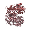
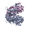
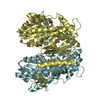
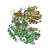
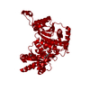
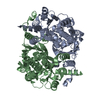
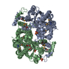
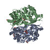
| Entry | Database: PDB / ID: 7nss | ||||||
|---|---|---|---|---|---|---|---|
| Title | Bdellovibrio bacteriovorus PGI in P3121 spacegroup | ||||||
 Components Components | Glucose-6-phosphate isomerase | ||||||
 Keywords Keywords | ISOMERASE / Glucose-6-phosphate isomerase Isomerase Phosphoglucose isomerase | ||||||
| Function / homology |  Function and homology informationglucose-6-phosphate isomerase / glucose-6-phosphate isomerase activity / glucose 6-phosphate metabolic process / carbohydrate derivative binding / monosaccharide binding / gluconeogenesis / glycolytic process / cytosol Function and homology informationglucose-6-phosphate isomerase / glucose-6-phosphate isomerase activity / glucose 6-phosphate metabolic process / carbohydrate derivative binding / monosaccharide binding / gluconeogenesis / glycolytic process / cytosolSimilarity search - Function | ||||||
| Biological species |  Bdellovibrio bacteriovorus (bacteria) Bdellovibrio bacteriovorus (bacteria) | ||||||
| Method | X-RAY DIFFRACTION / SYNCHROTRON / MOLECULAR REPLACEMENT / Resolution: 1.84 Å | ||||||
 Authors Authors | Meek, R.W. / Lovering, A.L. | ||||||
| Funding support |  United Kingdom, 1items United Kingdom, 1items
| ||||||
 Citation Citation | Journal: Open Biology / Year: 2021 Title: Bdellovibrio bacteriovorus phosphoglucose isomerase structures reveal novel rigidity in the active site of a selected subset of enzymes upon substrate binding. Authors: Meek, R.W. / Cadby, I.T. / Lovering, A.L. | ||||||
| History |
|
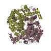
- Structure visualization
| Structure viewer | Molecule: MolmilJmol/JSmol |
|---|
- Downloads & links
-Download
| PDBx/mmCIF format | 7nss.cif.gz | 98.8 KB | Display | PDBx/mmCIF format |
|---|---|---|---|---|
| PDB format | pdb7nss.ent.gz | 74.1 KB | Display | PDB format |
| PDBx/mmJSON format | 7nss.json.gz | Tree view | PDBx/mmJSON format | |
| Others |  Other downloads Other downloads |
-Validation report
| Arichive directory | https://data.pdbj.org/pub/pdb/validation_reports/ns/7nssftp://data.pdbj.org/pub/pdb/validation_reports/ns/7nss | HTTPS FTP |
|---|
-Related structure data
| Related structure data |  7ntgC  7o0aC  1b0zS S: Starting model for refinement C: citing same article ( |
|---|---|
| Similar structure data |

-Links
 PDBj
PDBj

- Assembly
| Deposited unit | 
| ||||||||
|---|---|---|---|---|---|---|---|---|---|
| 1 | 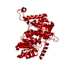
| ||||||||
| Unit cell |
|
-Components
| #1: Protein | Mass: 48055.336 Da / Num. of mol.: 1 Source method: isolated from a genetically manipulated source Source: (gene. exp.) Bdellovibrio bacteriovorus (strain ATCC 15356 / DSM 50701 / NCIB 9529 / HD100) (bacteria)Strain: ATCC 15356 / DSM 50701 / NCIB 9529 / HD100 / Gene: pgi, Bd0741 / Production host: Escherichia coli BL21(DE3) (bacteria) / References: UniProt: Q6MPU9, glucose-6-phosphate isomerase |
|---|---|
| #2: Chemical | ChemComp-EDO / Ethylene glycol  Mass: 62.068 Da / Num. of mol.: 1 / Source method: obtained synthetically / Formula: C2H6O2 Mass: 62.068 Da / Num. of mol.: 1 / Source method: obtained synthetically / Formula: C2H6O2 |
| #3: Water | ChemComp-HOH / Water Mass: 18.015 Da / Num. of mol.: 249 / Source method: isolated from a natural source / Formula: H2O Mass: 18.015 Da / Num. of mol.: 249 / Source method: isolated from a natural source / Formula: H2O |
| Has ligand of interest | N |
-Experimental details
-Experiment
| Experiment | Method: X-RAY DIFFRACTION / Number of used crystals: 1 |
|---|
- Sample preparation
| Crystal | Density Matthews: 2.35 Å3/Da / Density % sol: 47.69 % |
|---|---|
| Crystal grow | Temperature: 291 K / Method: vapor diffusion, sitting drop / pH: 5 Details: 0.2 M ammonium acetate, 0.1 M sodium acetate pH 5.0 and 20% PEG 4000 |
-Data collection
| Diffraction | Mean temperature: 100 K / Serial crystal experiment: N |
|---|---|
| Diffraction source | Source: SYNCHROTRON / Site: Diamond / Beamline: I04 / Wavelength: 0.9795 Å |
| Detector | Type: DECTRIS PILATUS 6M-F / Detector: PIXEL / Date: Jun 8, 2016 |
| Radiation | Protocol: SINGLE WAVELENGTH / Monochromatic (M) / Laue (L): M / Scattering type: x-ray |
| Radiation wavelength | Wavelength: 0.9795 Å / Relative weight: 1 |
| Reflection | Resolution: 1.84→57.74 Å / Num. obs: 37920 / % possible obs: 95.8 % / Redundancy: 18.7 % / Biso Wilson estimate: 28.46 Å2 / CC1/2: 1 / Rmerge(I) obs: 0.101 / Rpim(I) all: 0.034 / Rrim(I) all: 0.106 / Net I/σ(I): 23.2 |
| Reflection shell | Resolution: 1.84→1.87 Å / Rmerge(I) obs: 1.919 / Mean I/σ(I) obs: 1.3 / Num. unique obs: 2876 / CC1/2: 0.559 / Rpim(I) all: 0.821 |
- Processing
| Software |
| ||||||||||||||||||||||||||||||||||||||||||||||||||||||||||||
|---|---|---|---|---|---|---|---|---|---|---|---|---|---|---|---|---|---|---|---|---|---|---|---|---|---|---|---|---|---|---|---|---|---|---|---|---|---|---|---|---|---|---|---|---|---|---|---|---|---|---|---|---|---|---|---|---|---|---|---|---|---|
| Refinement | Method to determine structure: MOLECULAR REPLACEMENT Starting model: 1b0z Resolution: 1.84→57.74 Å / Cor.coef. Fo:Fc: 0.964 / Cor.coef. Fo:Fc free: 0.955 / Cross valid method: THROUGHOUT / σ(F): 0 / ESU R: 0.141 / ESU R Free: 0.129 / Stereochemistry target values: MAXIMUM LIKELIHOOD Details: HYDROGENS HAVE BEEN ADDED IN THE RIDING POSITIONS U VALUES : REFINED INDIVIDUALLY
| ||||||||||||||||||||||||||||||||||||||||||||||||||||||||||||
| Solvent computation | Ion probe radii: 0.8 Å / Shrinkage radii: 0.8 Å / VDW probe radii: 1.2 Å / Solvent model: MASK | ||||||||||||||||||||||||||||||||||||||||||||||||||||||||||||
| Displacement parameters | Biso max: 98.86 Å2 / Biso mean: 35.017 Å2 / Biso min: 21.06 Å2
| ||||||||||||||||||||||||||||||||||||||||||||||||||||||||||||
| Refinement step | Cycle: final / Resolution: 1.84→57.74 Å
| ||||||||||||||||||||||||||||||||||||||||||||||||||||||||||||
| Refine LS restraints |
| ||||||||||||||||||||||||||||||||||||||||||||||||||||||||||||
| LS refinement shell | Resolution: 1.84→1.888 Å / Rfactor Rfree error: 0 / Total num. of bins used: 20
|
