 Movie
Movie Controller

[English] 日本語
 Yorodumi
- PDB-7ns7: Human L-alanine:glyoxylate aminotransferase minor allele variant:... -
+ Open data

- Basic information
| Entry | Database: PDB / ID: 7ns7 | ||||||
|---|---|---|---|---|---|---|---|
| Title | Human L-alanine:glyoxylate aminotransferase minor allele variant: AGXT-Mi (P11L-I340M) | ||||||
 Components Components | L-alanine:glyoxylate aminotransferase | ||||||
 Keywords Keywords |  TRANSFERASE / alanine-glyoxylate transaminase / alanine-glyoxylate aminotransferase / alanine-glyoxylic aminotransferase / L-alanine-glycine transaminase / pyrodoxal / disorder TRANSFERASE / alanine-glyoxylate transaminase / alanine-glyoxylate aminotransferase / alanine-glyoxylic aminotransferase / L-alanine-glycine transaminase / pyrodoxal / disorder | ||||||
| Function / homology |  Function and homology information Function and homology informationoxalic acid secretion / serine-pyruvate transaminase / alanine-glyoxylate transaminase / glycine biosynthetic process, by transamination of glyoxylate / glyoxylate metabolic process / serine-pyruvate transaminase activity / L-alanine catabolic process / alanine-glyoxylate transaminase activity / L-serine metabolic process / L-cysteine catabolic process ...oxalic acid secretion / serine-pyruvate transaminase / alanine-glyoxylate transaminase / glycine biosynthetic process, by transamination of glyoxylate / glyoxylate metabolic process / serine-pyruvate transaminase activity / L-alanine catabolic process / alanine-glyoxylate transaminase activity / L-serine metabolic process / L-cysteine catabolic process / glyoxylate catabolic process / Glyoxylate metabolism and glycine degradation / transaminase activity / amino acid binding / peroxisomal matrix / Notch signaling pathway / Peroxisomal protein import / peroxisome / : / pyridoxal phosphate binding / intracellular membrane-bounded organelle / protein homodimerization activity / identical protein binding / cytosolSimilarity search - Function | ||||||
| Biological species |  Homo sapiens (human) Homo sapiens (human) | ||||||
| Method | X-RAY DIFFRACTION / SYNCHROTRON / MOLECULAR REPLACEMENT / molecular replacement / Resolution: 2.2 Å | ||||||
 Authors Authors | Cellini, B. / Dindo, M. | ||||||
 Citation Citation | Journal: Protein Sci. / Year: 2022 Title: Structural dynamics shape the fitness window of alanine:glyoxylate aminotransferase. Authors: Dindo, M. / Pascarelli, S. / Chiasserini, D. / Grottelli, S. / Costantini, C. / Uechi, G.I. / Giardina, G. / Laurino, P. / Cellini, B. | ||||||
| History |
|
- Structure visualization
| Structure viewer | Molecule: MolmilJmol/JSmol |
|---|
- Downloads & links
-Download
| PDBx/mmCIF format | 7ns7.cif.gz | 144 KB | Display | PDBx/mmCIF format |
|---|---|---|---|---|
| PDB format | pdb7ns7.ent.gz | 111.1 KB | Display | PDB format |
| PDBx/mmJSON format | 7ns7.json.gz | Tree view | PDBx/mmJSON format | |
| Others |  Other downloads Other downloads |
-Validation report
| Arichive directory | https://data.pdbj.org/pub/pdb/validation_reports/ns/7ns7ftp://data.pdbj.org/pub/pdb/validation_reports/ns/7ns7 | HTTPS FTP |
|---|
-Related structure data
| Related structure data | 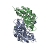 5f9sS S: Starting model for refinement |
|---|---|
| Similar structure data |
-Links
 PDBj
PDBj- Assembly
| Deposited unit | 
| ||||||||
|---|---|---|---|---|---|---|---|---|---|
| 1 |
| ||||||||
| Unit cell |
|
-Components
| #1: Protein | Mass: 43325.039 Da / Num. of mol.: 2 Source method: isolated from a genetically manipulated source Details: polymorphic variant P11L-I340M. PLP bound to K209 internal aldimine form Source: (gene. exp.) Homo sapiens (human) / Gene: AGXT / Variant: P11L / Production host:  Escherichia coli (E. coli) Escherichia coli (E. coli)References: UniProt: P21549, alanine-glyoxylate transaminase, serine-pyruvate transaminase#2: Water | ChemComp-HOH / | Water Mass: 18.015 Da / Num. of mol.: 75 / Source method: isolated from a natural source / Formula: H2O Mass: 18.015 Da / Num. of mol.: 75 / Source method: isolated from a natural source / Formula: H2OHas ligand of interest | N | |
|---|
-Experimental details
-Experiment
| Experiment | Method: X-RAY DIFFRACTION / Number of used crystals: 1 |
|---|
- Sample preparation
| Crystal | Density Matthews: 3.34 Å3/Da / Density % sol: 63.12 % / Description: diamond shaped yellow crystals |
|---|---|
| Crystal grow | Temperature: 294 K / Method: vapor diffusion, sitting drop / pH: 7.5 Details: 1:1 mixing of: Protein: 0.04M AGT-Mi solution in 0.1M KPhO Precipitant: 0.03M MgCl2 , 0.03M CaCl2 , 0.05M HEPES 0.05M MOPS at a final pH 7.5, 40% v/v Ethylene glycol; 20% w/v PEG 8K |
-Data collection
| Diffraction | Mean temperature: 100 K / Serial crystal experiment: N | ||||||||||||||||||||||||||||||
|---|---|---|---|---|---|---|---|---|---|---|---|---|---|---|---|---|---|---|---|---|---|---|---|---|---|---|---|---|---|---|---|
| Diffraction source | Source: SYNCHROTRON / Site: ELETTRA  / Beamline: 5.2R / Wavelength: 1 Å / Beamline: 5.2R / Wavelength: 1 Å | ||||||||||||||||||||||||||||||
| Detector | Type: DECTRIS PILATUS 2M / Detector: PIXEL / Date: Nov 8, 2018 | ||||||||||||||||||||||||||||||
| Radiation | Monochromator: Si(111) / Protocol: SINGLE WAVELENGTH / Monochromatic (M) / Laue (L): M / Scattering type: x-ray | ||||||||||||||||||||||||||||||
| Radiation wavelength | Wavelength: 1 Å / Relative weight: 1 | ||||||||||||||||||||||||||||||
| Reflection twin |
| ||||||||||||||||||||||||||||||
| Reflection | Resolution: 2.2→64.06 Å / Num. obs: 57290 / % possible obs: 99.6 % / Redundancy: 3.8 % / CC1/2: 0.992 / Rmerge(I) obs: 0.109 / Rpim(I) all: 0.061 / Rrim(I) all: 0.125 / Net I/σ(I): 9.3 / Num. measured all: 214921 / Scaling rejects: 58 | ||||||||||||||||||||||||||||||
| Reflection shell | Diffraction-ID: 1
|
-Phasing
| Phasing | Method: molecular replacement |
|---|
- Processing
| Software |
| ||||||||||||||||||||||||||||||||||||||||||||||||||||||||||||
|---|---|---|---|---|---|---|---|---|---|---|---|---|---|---|---|---|---|---|---|---|---|---|---|---|---|---|---|---|---|---|---|---|---|---|---|---|---|---|---|---|---|---|---|---|---|---|---|---|---|---|---|---|---|---|---|---|---|---|---|---|---|
| Refinement | Method to determine structure: MOLECULAR REPLACEMENT Starting model: 5F9S Resolution: 2.2→64.06 Å / Cor.coef. Fo:Fc: 0.887 / Cor.coef. Fo:Fc free: 0.831 / SU B: 5.292 / SU ML: 0.139 / Cross valid method: THROUGHOUT / σ(F): 0 / ESU R: 0.048 / ESU R Free: 0.044 / Stereochemistry target values: MAXIMUM LIKELIHOOD Details: HYDROGENS HAVE BEEN ADDED IN THE RIDING POSITIONS U VALUES : REFINED INDIVIDUALLY
| ||||||||||||||||||||||||||||||||||||||||||||||||||||||||||||
| Solvent computation | Ion probe radii: 0.8 Å / Shrinkage radii: 0.8 Å / VDW probe radii: 1.2 Å / Solvent model: MASK | ||||||||||||||||||||||||||||||||||||||||||||||||||||||||||||
| Displacement parameters | Biso max: 78.54 Å2 / Biso mean: 26.561 Å2 / Biso min: 5.95 Å2
| ||||||||||||||||||||||||||||||||||||||||||||||||||||||||||||
| Refinement step | Cycle: final / Resolution: 2.2→64.06 Å
| ||||||||||||||||||||||||||||||||||||||||||||||||||||||||||||
| Refine LS restraints |
| ||||||||||||||||||||||||||||||||||||||||||||||||||||||||||||
| LS refinement shell | Resolution: 2.2→2.257 Å / Rfactor Rfree error: 0 / Total num. of bins used: 20
|
