 Movie
Movie Controller

[English] 日本語
 Yorodumi
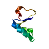
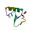
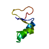
- PDB-7npq: Crystal structure of the human LL37(17-29) I24C mutant antimicrob... -
+ Open data

- Basic information
| Entry | Database: PDB / ID: 7npq | ||||||
|---|---|---|---|---|---|---|---|
| Title | Crystal structure of the human LL37(17-29) I24C mutant antimicrobial peptide | ||||||
 Components Components | Cathelicidin antimicrobial peptide | ||||||
 Keywords Keywords |  ANTIMICROBIAL PROTEIN / LL-37 / functional fibril / helical fibril / AMPs / cys mutant / design / ANTIMICROBIAL peptide ANTIMICROBIAL PROTEIN / LL-37 / functional fibril / helical fibril / AMPs / cys mutant / design / ANTIMICROBIAL peptide | ||||||
| Function / homology |  Function and homology informationcytolysis / killing by host of symbiont cells / neutrophil activation / specific granule / cellular response to peptidoglycan / cellular response to interleukin-6 / Antimicrobial peptides / cellular response to interleukin-1 / innate immune response in mucosa / cell projection ...cytolysis / killing by host of symbiont cells / neutrophil activation / specific granule / cellular response to peptidoglycan / cellular response to interleukin-6 / Antimicrobial peptides / cellular response to interleukin-1 / innate immune response in mucosa / cell projection / lipopolysaccharide binding / specific granule lumen / positive regulation of angiogenesis / antimicrobial humoral immune response mediated by antimicrobial peptide / tertiary granule lumen / cellular response to tumor necrosis factor / antibacterial humoral response / cellular response to lipopolysaccharide / defense response to Gram-negative bacterium / amyloid fibril formation / defense response to Gram-positive bacterium / defense response to bacterium / positive regulation of protein phosphorylation / innate immune response / positive regulation of cell population proliferation / Neutrophil degranulation / extracellular space / extracellular exosome / extracellular region Function and homology informationcytolysis / killing by host of symbiont cells / neutrophil activation / specific granule / cellular response to peptidoglycan / cellular response to interleukin-6 / Antimicrobial peptides / cellular response to interleukin-1 / innate immune response in mucosa / cell projection ...cytolysis / killing by host of symbiont cells / neutrophil activation / specific granule / cellular response to peptidoglycan / cellular response to interleukin-6 / Antimicrobial peptides / cellular response to interleukin-1 / innate immune response in mucosa / cell projection / lipopolysaccharide binding / specific granule lumen / positive regulation of angiogenesis / antimicrobial humoral immune response mediated by antimicrobial peptide / tertiary granule lumen / cellular response to tumor necrosis factor / antibacterial humoral response / cellular response to lipopolysaccharide / defense response to Gram-negative bacterium / amyloid fibril formation / defense response to Gram-positive bacterium / defense response to bacterium / positive regulation of protein phosphorylation / innate immune response / positive regulation of cell population proliferation / Neutrophil degranulation / extracellular space / extracellular exosome / extracellular regionSimilarity search - Function | ||||||
| Biological species |  Homo sapiens (human) Homo sapiens (human) | ||||||
| Method | X-RAY DIFFRACTION / SYNCHROTRON / MOLECULAR REPLACEMENT / molecular replacement / Resolution: 1.5 Å | ||||||
| Model details | Antimicrobial | ||||||
 Authors Authors | Landau, M. / Engelberg, Y. | ||||||
 Citation Citation | Journal: Biomacromolecules / Year: 2022 Title: Rare by Natural Selection: Disulfide-Bonded Supramolecular Antimicrobial Peptides. Authors: Engelberg, Y. / Ragonis-Bachar, P. / Landau, M. | ||||||
| History |
|
- Structure visualization
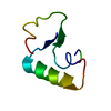
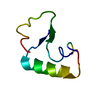
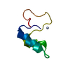
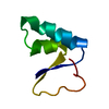
| Structure viewer | Molecule: MolmilJmol/JSmol |
|---|
- Downloads & links
-Download
| PDBx/mmCIF format | 7npq.cif.gz | 17.8 KB | Display | PDBx/mmCIF format |
|---|---|---|---|---|
| PDB format | pdb7npq.ent.gz | 10.1 KB | Display | PDB format |
| PDBx/mmJSON format | 7npq.json.gz | Tree view | PDBx/mmJSON format | |
| Others |  Other downloads Other downloads |
-Validation report
| Arichive directory | https://data.pdbj.org/pub/pdb/validation_reports/np/7npqftp://data.pdbj.org/pub/pdb/validation_reports/np/7npq | HTTPS FTP |
|---|
-Related structure data
| Related structure data |  6s6mS S: Starting model for refinement |
|---|---|
| Similar structure data |


-Links
 PDBj
PDBj

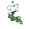
- Assembly
| Deposited unit | 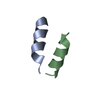
| ||||||||
|---|---|---|---|---|---|---|---|---|---|
| 1 |
| ||||||||
| Unit cell |
| ||||||||
| Components on special symmetry positions |
|
-Components
| #1: Protein/peptide | Mass: 1714.109 Da / Num. of mol.: 2 Fragment: Antimicrobial core segment of human LL37 (residues 17-29) I8C mutant Source method: obtained synthetically / Details: Human LL-37(17-29) I24C mutant, synthesized / Source: (synth.) Homo sapiens (human) / References: UniProt: P49913#2: Water | ChemComp-HOH / | Water Mass: 18.015 Da / Num. of mol.: 16 / Source method: isolated from a natural source / Formula: H2O Mass: 18.015 Da / Num. of mol.: 16 / Source method: isolated from a natural source / Formula: H2O |
|---|
-Experimental details
-Experiment
| Experiment | Method: X-RAY DIFFRACTION / Number of used crystals: 1 |
|---|
- Sample preparation
| Crystal | Density Matthews: 1.98 Å3/Da / Density % sol: 37.93 % |
|---|---|
| Crystal grow | Temperature: 293 K / Method: vapor diffusion, hanging drop Details: The LL-37(17-29) I24C peptide was mixed with 0.1mM DTT in water: Reservoir contained 2.8 M sodium acetate trihydrate pH 7.0 |
-Data collection
| Diffraction | Mean temperature: 100 K / Serial crystal experiment: N | ||||||||||||||||||||||||||||||||||||||||||||||||||||||||||||||||||||||||||||||||||||||||||||||||||||||||||||||
|---|---|---|---|---|---|---|---|---|---|---|---|---|---|---|---|---|---|---|---|---|---|---|---|---|---|---|---|---|---|---|---|---|---|---|---|---|---|---|---|---|---|---|---|---|---|---|---|---|---|---|---|---|---|---|---|---|---|---|---|---|---|---|---|---|---|---|---|---|---|---|---|---|---|---|---|---|---|---|---|---|---|---|---|---|---|---|---|---|---|---|---|---|---|---|---|---|---|---|---|---|---|---|---|---|---|---|---|---|---|---|---|
| Diffraction source | Source: SYNCHROTRON / Site: PETRA III, EMBL c/o DESY  / Beamline: P14 (MX2) / Wavelength: 0.9763 Å / Beamline: P14 (MX2) / Wavelength: 0.9763 Å | ||||||||||||||||||||||||||||||||||||||||||||||||||||||||||||||||||||||||||||||||||||||||||||||||||||||||||||||
| Detector | Type: DECTRIS EIGER X 16M / Detector: PIXEL / Date: Dec 17, 2019 | ||||||||||||||||||||||||||||||||||||||||||||||||||||||||||||||||||||||||||||||||||||||||||||||||||||||||||||||
| Radiation | Protocol: SINGLE WAVELENGTH / Monochromatic (M) / Laue (L): M / Scattering type: x-ray | ||||||||||||||||||||||||||||||||||||||||||||||||||||||||||||||||||||||||||||||||||||||||||||||||||||||||||||||
| Radiation wavelength | Wavelength: 0.9763 Å / Relative weight: 1 | ||||||||||||||||||||||||||||||||||||||||||||||||||||||||||||||||||||||||||||||||||||||||||||||||||||||||||||||
| Reflection | Resolution: 1.5→27.49 Å / Num. obs: 4721 / % possible obs: 99 % / Redundancy: 7.571 % / Biso Wilson estimate: 29.339 Å2 / CC1/2: 0.999 / Rmerge(I) obs: 0.052 / Rrim(I) all: 0.056 / Χ2: 1.042 / Net I/σ(I): 19.87 / Num. measured all: 35742 | ||||||||||||||||||||||||||||||||||||||||||||||||||||||||||||||||||||||||||||||||||||||||||||||||||||||||||||||
| Reflection shell | Diffraction-ID: 1
|
-Phasing
| Phasing | Method: molecular replacement |
|---|---|
| Phasing MR | Packing: 0 |
- Processing
| Software |
| ||||||||||||||||||||||||||||||||||||||||||||||||||||||||||||
|---|---|---|---|---|---|---|---|---|---|---|---|---|---|---|---|---|---|---|---|---|---|---|---|---|---|---|---|---|---|---|---|---|---|---|---|---|---|---|---|---|---|---|---|---|---|---|---|---|---|---|---|---|---|---|---|---|---|---|---|---|---|
| Refinement | Method to determine structure: MOLECULAR REPLACEMENT Starting model: PDBid 6S6M with residues mutated to alanine Resolution: 1.5→27.49 Å / Cor.coef. Fo:Fc: 0.962 / Cor.coef. Fo:Fc free: 0.962 / WRfactor Rfree: 0.1948 / WRfactor Rwork: 0.1894 / FOM work R set: 0.8614 / SU B: 1.354 / SU ML: 0.049 / SU R Cruickshank DPI: 0.082 / SU Rfree: 0.0757 / Cross valid method: THROUGHOUT / σ(F): 0 / ESU R: 0.082 / ESU R Free: 0.076 / Stereochemistry target values: MAXIMUM LIKELIHOOD Details: HYDROGENS HAVE BEEN ADDED IN THE RIDING POSITIONS U VALUES : REFINED INDIVIDUALLY
| ||||||||||||||||||||||||||||||||||||||||||||||||||||||||||||
| Solvent computation | Ion probe radii: 0.8 Å / Shrinkage radii: 0.8 Å / VDW probe radii: 1.2 Å / Solvent model: MASK | ||||||||||||||||||||||||||||||||||||||||||||||||||||||||||||
| Displacement parameters | Biso max: 83 Å2 / Biso mean: 28.058 Å2 / Biso min: 15.06 Å2
| ||||||||||||||||||||||||||||||||||||||||||||||||||||||||||||
| Refinement step | Cycle: final / Resolution: 1.5→27.49 Å
| ||||||||||||||||||||||||||||||||||||||||||||||||||||||||||||
| Refine LS restraints |
| ||||||||||||||||||||||||||||||||||||||||||||||||||||||||||||
| LS refinement shell | Resolution: 1.5→1.539 Å / Rfactor Rfree error: 0 / Total num. of bins used: 20
|
