 Movie
Movie Controller

[English] 日本語
 Yorodumi
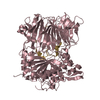
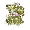
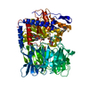
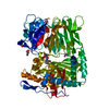
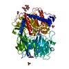
- PDB-7ne4: E125A mutant of oligopeptidase B from S. proteomaculans with modi... -
+ Open data

- Basic information
| Entry | Database: PDB / ID: 7ne4 | ||||||
|---|---|---|---|---|---|---|---|
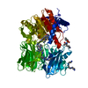
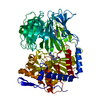
| Title | E125A mutant of oligopeptidase B from S. proteomaculans with modified hinge region | ||||||
 Components Components | Oligopeptidase B | ||||||
 Keywords Keywords | HYDROLASE | ||||||
| Function / homology |  Function and homology information Function and homology information | ||||||
| Biological species |  Serratia proteamaculans (bacteria) Serratia proteamaculans (bacteria) | ||||||
| Method | X-RAY DIFFRACTION / SYNCHROTRON / MOLECULAR REPLACEMENT / Resolution: 2.717 Å | ||||||
 Authors Authors | Petrenko, D.E. / Nikolaeva, A.Y. / Lazarenko, V.A. / Dorovatovskiy, P.V. / Vlaskina, A.V. / Mikhailova, A.G. / Rakitina, T.V. / Timofeev, V.I. | ||||||
 Citation Citation | Journal: Biology (Basel) / Year: 2021 Title: First Crystal Structure of Bacterial Oligopeptidase B in an Intermediate State: The Roles of the Hinge Region Modification and Spermine. Authors: Petrenko, D.E. / Timofeev, V.I. / Britikov, V.V. / Britikova, E.V. / Kleymenov, S.Y. / Vlaskina, A.V. / Kuranova, I.P. / Mikhailova, A.G. / Rakitina, T.V. | ||||||
| History |
|
- Structure visualization
| Structure viewer | Molecule: MolmilJmol/JSmol |
|---|
- Downloads & links
-Download
| PDBx/mmCIF format | 7ne4.cif.gz | 148 KB | Display | PDBx/mmCIF format |
|---|---|---|---|---|
| PDB format | pdb7ne4.ent.gz | 113.8 KB | Display | PDB format |
| PDBx/mmJSON format | 7ne4.json.gz | Tree view | PDBx/mmJSON format | |
| Others |  Other downloads Other downloads |
-Validation report
| Arichive directory | https://data.pdbj.org/pub/pdb/validation_reports/ne/7ne4ftp://data.pdbj.org/pub/pdb/validation_reports/ne/7ne4 | HTTPS FTP |
|---|
-Related structure data
| Related structure data |  7ne5C 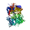 7ob1C  6tf5 S: Starting model for refinement C: citing same article ( |
|---|---|
| Similar structure data |

-Links
 PDBj
PDBj


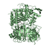
- Assembly
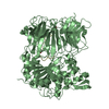
| Deposited unit | 
| ||||||||
|---|---|---|---|---|---|---|---|---|---|
| 1 |
| ||||||||
| Unit cell |
|
-Components
| #1: Protein | Mass: 78263.664 Da / Num. of mol.: 1 Source method: isolated from a genetically manipulated source Source: (gene. exp.) Serratia proteamaculans (bacteria) / Gene: opdB / Production host: Escherichia coli (E. coli) / References: UniProt: B3VI58 | ||||
|---|---|---|---|---|---|
| #2: Chemical | Spermine  Mass: 202.340 Da / Num. of mol.: 3 / Source method: obtained synthetically / Formula: C10H26N4 Mass: 202.340 Da / Num. of mol.: 3 / Source method: obtained synthetically / Formula: C10H26N4#3: Water | ChemComp-HOH / | Water Mass: 18.015 Da / Num. of mol.: 50 / Source method: isolated from a natural source / Formula: H2O Mass: 18.015 Da / Num. of mol.: 50 / Source method: isolated from a natural source / Formula: H2OHas ligand of interest | N | |
-Experimental details
-Experiment
| Experiment | Method: X-RAY DIFFRACTION / Number of used crystals: 1 |
|---|
- Sample preparation
| Crystal | Density Matthews: 2.35 Å3/Da / Density % sol: 47.58 % |
|---|---|
| Crystal grow | Temperature: 277 K / Method: vapor diffusion, hanging drop Details: 200 mM Lithium sulfate, 100 mM Bis-Tris pH 5.5, 23% PEG 3350 |
-Data collection
| Diffraction | Mean temperature: 100 K / Serial crystal experiment: N |
|---|---|
| Diffraction source | Source: SYNCHROTRON / Site: KURCHATOV SNC  / Beamline: K4.4 / Wavelength: 0.8 Å / Beamline: K4.4 / Wavelength: 0.8 Å |
| Detector | Type: RAYONIX MX-225 / Detector: CCD / Date: Mar 12, 2019 |
| Radiation | Protocol: SINGLE WAVELENGTH / Monochromatic (M) / Laue (L): M / Scattering type: x-ray |
| Radiation wavelength | Wavelength: 0.8 Å / Relative weight: 1 |
| Reflection | Resolution: 2.717→44.9 Å / Num. obs: 20453 / % possible obs: 99.92 % / Redundancy: 6.18 % / Rmerge(I) obs: 0.061 / Net I/σ(I): 8.45 |
| Reflection shell | Resolution: 2.717→2.79 Å / Rmerge(I) obs: 0.29 / Mean I/σ(I) obs: 2.11 / Num. unique obs: 1476 |
- Processing
| Software |
| ||||||||||||||||||||||||||||||||||||||||||||||||||||||||||||
|---|---|---|---|---|---|---|---|---|---|---|---|---|---|---|---|---|---|---|---|---|---|---|---|---|---|---|---|---|---|---|---|---|---|---|---|---|---|---|---|---|---|---|---|---|---|---|---|---|---|---|---|---|---|---|---|---|---|---|---|---|---|
| Refinement | Method to determine structure: MOLECULAR REPLACEMENT Starting model: 6TF5 6tf5 Resolution: 2.717→44.85 Å / Cor.coef. Fo:Fc: 0.894 / Cor.coef. Fo:Fc free: 0.829 / SU B: 44.184 / SU ML: 0.412 / Cross valid method: THROUGHOUT / σ(F): 0 / ESU R Free: 0.455 / Stereochemistry target values: MAXIMUM LIKELIHOOD Details: HYDROGENS HAVE BEEN ADDED IN THE RIDING POSITIONS U VALUES : RESIDUAL ONLY
| ||||||||||||||||||||||||||||||||||||||||||||||||||||||||||||
| Solvent computation | Ion probe radii: 0.8 Å / Shrinkage radii: 0.8 Å / VDW probe radii: 1.1 Å / Solvent model: MASK | ||||||||||||||||||||||||||||||||||||||||||||||||||||||||||||
| Displacement parameters | Biso max: 65.54 Å2 / Biso mean: 28.828 Å2 / Biso min: 10.18 Å2
| ||||||||||||||||||||||||||||||||||||||||||||||||||||||||||||
| Refinement step | Cycle: final / Resolution: 2.717→44.85 Å
| ||||||||||||||||||||||||||||||||||||||||||||||||||||||||||||
| Refine LS restraints |
| ||||||||||||||||||||||||||||||||||||||||||||||||||||||||||||
| LS refinement shell | Resolution: 2.717→2.788 Å / Rfactor Rfree error: 0
|
