 Movie
Movie Controller


+ Open data

- Basic information
| Entry | Database: PDB / ID: 7mhx | ||||||
|---|---|---|---|---|---|---|---|
| Title | KcsA E71V closed gate with Ba2+ | ||||||
 Components Components |
| ||||||
 Keywords Keywords |  MEMBRANE PROTEIN / ion channel MEMBRANE PROTEIN / ion channel | ||||||
| Function / homology | Potassium channel domain / Ion channel / monoatomic ion transmembrane transport / identical protein binding / plasma membrane / : / DIACYL GLYCEROL / pH-gated potassium channel KcsA Function and homology information Function and homology information | ||||||
| Biological species |  Mus musculus (house mouse) Mus musculus (house mouse) Streptomyces lividans (bacteria) Streptomyces lividans (bacteria) | ||||||
| Method | X-RAY DIFFRACTION / SYNCHROTRON / MOLECULAR REPLACEMENT / Resolution: 2.85 Å | ||||||
 Authors Authors | Rohaim, A. / Li, J. / Weingarth, M. / Roux, B. | ||||||
| Funding support |  United States, 1items United States, 1items
| ||||||
 Citation Citation | Journal: Nat Commun / Year: 2022 Title: A distinct mechanism of C-type inactivation in the Kv-like KcsA mutant E71V. Authors: Rohaim, A. / Vermeulen, B.J.A. / Li, J. / Kummerer, F. / Napoli, F. / Blachowicz, L. / Medeiros-Silva, J. / Roux, B. / Weingarth, M. | ||||||
| History |
|
- Structure visualization
| Structure viewer | Molecule: MolmilJmol/JSmol |
|---|
- Downloads & links
-Download
| PDBx/mmCIF format | 7mhx.cif.gz | 220.7 KB | Display | PDBx/mmCIF format |
|---|---|---|---|---|
| PDB format | pdb7mhx.ent.gz | 176.9 KB | Display | PDB format |
| PDBx/mmJSON format | 7mhx.json.gz | Tree view | PDBx/mmJSON format | |
| Others |  Other downloads Other downloads |
-Validation report
| Arichive directory | https://data.pdbj.org/pub/pdb/validation_reports/mh/7mhxftp://data.pdbj.org/pub/pdb/validation_reports/mh/7mhx | HTTPS FTP |
|---|
-Related structure data
| Related structure data |  7mhrC 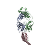 7mjtC 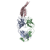 7mk6C 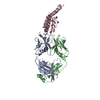 7mubC  1k4cS C: citing same article ( S: Starting model for refinement |
|---|---|
| Similar structure data |
-Links
 PDBj
PDBj


- Assembly
| Deposited unit | 
| ||||||||||||
|---|---|---|---|---|---|---|---|---|---|---|---|---|---|
| 1 | 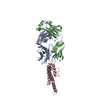
| ||||||||||||
| Unit cell |
| ||||||||||||
| Components on special symmetry positions |
|
-Components
-Protein , 1 types, 1 molecules C
| #3: Protein | Mass: 13181.600 Da / Num. of mol.: 1 / Mutation: P2A, E71V, L90C Source method: isolated from a genetically manipulated source Source: (gene. exp.) Streptomyces lividans (bacteria) / Gene: kcsA, skc1 / Production host: Escherichia coli (E. coli) / References: UniProt: P0A334 |
|---|
-Antibody , 2 types, 2 molecules AB
| #1: Antibody | Fragment antigen-binding Mass: 23411.242 Da / Num. of mol.: 1 Source method: isolated from a genetically manipulated source Source: (gene. exp.) Mus musculus (house mouse) / Production host: Mus musculus (house mouse) |
|---|---|
| #2: Antibody | Fragment antigen-binding Mass: 23435.738 Da / Num. of mol.: 1 Source method: isolated from a genetically manipulated source Source: (gene. exp.) Mus musculus (house mouse) / Production host: Mus musculus (house mouse) |
-Non-polymers , 3 types, 22 molecules 

| #4: Chemical | ChemComp-BA /  Mass: 137.327 Da / Num. of mol.: 1 / Source method: obtained synthetically / Formula: Ba / Feature type: SUBJECT OF INVESTIGATION Mass: 137.327 Da / Num. of mol.: 1 / Source method: obtained synthetically / Formula: Ba / Feature type: SUBJECT OF INVESTIGATION |
|---|---|
| #5: Chemical | ChemComp-DGA / Diglyceride Mass: 625.018 Da / Num. of mol.: 1 / Source method: isolated from a natural source / Formula: C39H76O5 / Feature type: SUBJECT OF INVESTIGATION Mass: 625.018 Da / Num. of mol.: 1 / Source method: isolated from a natural source / Formula: C39H76O5 / Feature type: SUBJECT OF INVESTIGATION |
| #6: Water | ChemComp-HOH / WaterMass: 18.015 Da / Num. of mol.: 20 / Source method: isolated from a natural source / Formula: H2O |
-Details
| Has ligand of interest | Y |
|---|
-Experimental details
-Experiment
| Experiment | Method: X-RAY DIFFRACTION / Number of used crystals: 1 |
|---|
- Sample preparation
| Crystal | Density Matthews: 3.94 Å3/Da / Density % sol: 68.76 % |
|---|---|
| Crystal grow | Temperature: 293 K / Method: vapor diffusion, hanging drop Details: 50 mM sodium acetate pH 5.5, 50 mM magnesium acetate, 25% PEG 400 |
-Data collection
| Diffraction | Mean temperature: 98 K / Serial crystal experiment: N |
|---|---|
| Diffraction source | Source: SYNCHROTRON / Site: APS / Beamline: 24-ID-E / Wavelength: 0.9792 Å |
| Detector | Type: DECTRIS EIGER X 16M / Detector: PIXEL / Date: Dec 3, 2019 |
| Radiation | Protocol: SINGLE WAVELENGTH / Monochromatic (M) / Laue (L): M / Scattering type: x-ray |
| Radiation wavelength | Wavelength: 0.9792 Å / Relative weight: 1 |
| Reflection | Resolution: 2.85→77.58 Å / Num. obs: 21076 / % possible obs: 99.8 % / Redundancy: 6 % / CC1/2: 0.92 / Net I/σ(I): 9 |
| Reflection shell | Resolution: 2.85→3 Å / Num. unique obs: 3082 / CC1/2: 0.14 |
- Processing
| Software |
| ||||||||||||||||||||||||||||||||||||||||||||||||||||||||||||||||||||||||||||||||||||||||||||||||||||
|---|---|---|---|---|---|---|---|---|---|---|---|---|---|---|---|---|---|---|---|---|---|---|---|---|---|---|---|---|---|---|---|---|---|---|---|---|---|---|---|---|---|---|---|---|---|---|---|---|---|---|---|---|---|---|---|---|---|---|---|---|---|---|---|---|---|---|---|---|---|---|---|---|---|---|---|---|---|---|---|---|---|---|---|---|---|---|---|---|---|---|---|---|---|---|---|---|---|---|---|---|---|
| Refinement | Method to determine structure: MOLECULAR REPLACEMENT Starting model: 1K4C Resolution: 2.85→50.01 Å / Cor.coef. Fo:Fc: 0.936 / Cor.coef. Fo:Fc free: 0.926 / SU B: 41.121 / SU ML: 0.333 / Cross valid method: THROUGHOUT / σ(F): 0 / ESU R: 0.615 / ESU R Free: 0.32 / Stereochemistry target values: MAXIMUM LIKELIHOOD Details: HYDROGENS HAVE BEEN ADDED IN THE RIDING POSITIONS U VALUES : WITH TLS ADDED
| ||||||||||||||||||||||||||||||||||||||||||||||||||||||||||||||||||||||||||||||||||||||||||||||||||||
| Solvent computation | Ion probe radii: 0.8 Å / Shrinkage radii: 0.8 Å / VDW probe radii: 1.2 Å / Solvent model: MASK | ||||||||||||||||||||||||||||||||||||||||||||||||||||||||||||||||||||||||||||||||||||||||||||||||||||
| Displacement parameters | Biso max: 260.04 Å2 / Biso mean: 70 Å2 / Biso min: 28.72 Å2
| ||||||||||||||||||||||||||||||||||||||||||||||||||||||||||||||||||||||||||||||||||||||||||||||||||||
| Refinement step | Cycle: final / Resolution: 2.85→50.01 Å
| ||||||||||||||||||||||||||||||||||||||||||||||||||||||||||||||||||||||||||||||||||||||||||||||||||||
| Refine LS restraints |
| ||||||||||||||||||||||||||||||||||||||||||||||||||||||||||||||||||||||||||||||||||||||||||||||||||||
| LS refinement shell | Resolution: 2.9→2.924 Å / Rfactor Rfree error: 0
| ||||||||||||||||||||||||||||||||||||||||||||||||||||||||||||||||||||||||||||||||||||||||||||||||||||
| Refinement TLS params. | Method: refined / Refine-ID: X-RAY DIFFRACTION
| ||||||||||||||||||||||||||||||||||||||||||||||||||||||||||||||||||||||||||||||||||||||||||||||||||||
| Refinement TLS group |
|
