 Movie
Movie Controller

[English] 日本語
 Yorodumi
- PDB-7mcs: Cryo-electron microscopy structure of TnsC(1-503)A225V bound to DNA -
+ Open data

- Basic information
| Entry | Database: PDB / ID: 7mcs | ||||||
|---|---|---|---|---|---|---|---|
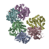
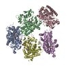
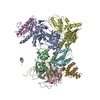
| Title | Cryo-electron microscopy structure of TnsC(1-503)A225V bound to DNA | ||||||
 Components Components |
| ||||||
 Keywords Keywords | DNA BINDING PROTEIN/DNA / AAA+ ATPase /  DNA binding / Transposition / Complex / DNA BINDING PROTEIN / DNA BINDING PROTEIN-DNA complex DNA binding / Transposition / Complex / DNA BINDING PROTEIN / DNA BINDING PROTEIN-DNA complex | ||||||
| Function / homology |  Function and homology information Function and homology informationtransposition / DNA recombination / ATP hydrolysis activity / DNA binding / ATP bindingSimilarity search - Function | ||||||
| Biological species |  Escherichia coli (E. coli) Escherichia coli (E. coli)synthetic construct (others) | ||||||
| Method | ELECTRON MICROSCOPY / single particle reconstruction / cryo EM / Resolution: 3.56 Å | ||||||
 Authors Authors | Shen, Y. / Ortega, J. / Guarne, A. | ||||||
| Funding support |  Canada, 1items Canada, 1items
| ||||||
 Citation Citation | Journal: Nat Struct Mol Biol / Year: 2022 Title: Structural basis for DNA targeting by the Tn7 transposon. Authors: Yao Shen / Josue Gomez-Blanco / Michael T Petassi / Joseph E Peters / Joaquin Ortega / Alba Guarné /  Abstract: Tn7 transposable elements are unique for their highly specific, and sometimes programmable, target-site selection mechanisms and precise insertions. All the elements in the Tn7 family utilize an AAA+ ...Tn7 transposable elements are unique for their highly specific, and sometimes programmable, target-site selection mechanisms and precise insertions. All the elements in the Tn7 family utilize an AAA+ adaptor (TnsC) to coordinate target-site selection with transpososome assembly and to prevent insertions at sites already containing a Tn7 element. Owing to its multiple functions, TnsC is considered the linchpin in the Tn7 element. Here we present the high-resolution cryo-EM structure of TnsC bound to DNA using a gain-of-function variant of the protein and a DNA substrate that together recapitulate the recruitment to a specific DNA target site. TnsC forms an asymmetric ring on target DNA that segregates target-site selection and interaction with the paired-end complex to opposite faces of the ring. Unlike most AAA+ ATPases, TnsC uses a DNA distortion to find the target site but does not remodel DNA to activate transposition. By recognizing pre-distorted substrates, TnsC creates a built-in regulatory mechanism where ATP hydrolysis abolishes ring formation proximal to an existing element. This work unveils how Tn7 and Tn7-like elements determine the strict spacing between the target and integration sites. | ||||||
| History |
|
- Structure visualization
| Movie |
Movie viewer |
|---|---|
| Structure viewer | Molecule: MolmilJmol/JSmol |
- Downloads & links
-Download
| PDBx/mmCIF format | 7mcs.cif.gz | 477.1 KB | Display | PDBx/mmCIF format |
|---|---|---|---|---|
| PDB format | pdb7mcs.ent.gz | 401 KB | Display | PDB format |
| PDBx/mmJSON format | 7mcs.json.gz | Tree view | PDBx/mmJSON format | |
| Others |  Other downloads Other downloads |
-Validation report
| Arichive directory | https://data.pdbj.org/pub/pdb/validation_reports/mc/7mcsftp://data.pdbj.org/pub/pdb/validation_reports/mc/7mcs | HTTPS FTP |
|---|
-Related structure data
| Related structure data |  23757MC  7mbwC M: map data used to model this data C: citing same article ( |
|---|---|
| Similar structure data |


-Links
 PDBj
PDBj


- Assembly
| Deposited unit | 
|
|---|---|
| 1 |
|
-Components
| #1: Protein | Mass: 59346.980 Da / Num. of mol.: 7 / Mutation: S2G, A225V Source method: isolated from a genetically manipulated source Source: (gene. exp.) Escherichia coli (E. coli) / Gene: tnsC / Production host: Escherichia coli (E. coli) / References: UniProt: P05846#2: DNA chain | | Mass: 4572.937 Da / Num. of mol.: 1 / Source method: obtained synthetically / Source: (synth.) synthetic construct (others) #3: DNA chain | | Mass: 4612.961 Da / Num. of mol.: 1 / Source method: obtained synthetically / Source: (synth.) synthetic construct (others) #4: Chemical | ChemComp-ANP /   Mass: 506.196 Da / Num. of mol.: 6 / Source method: obtained synthetically / Formula: C10H17N6O12P3 / Feature type: SUBJECT OF INVESTIGATION / Comment: AMP-PNP, energy-carrying molecule analogue*YM Mass: 506.196 Da / Num. of mol.: 6 / Source method: obtained synthetically / Formula: C10H17N6O12P3 / Feature type: SUBJECT OF INVESTIGATION / Comment: AMP-PNP, energy-carrying molecule analogue*YM#5: Chemical | ChemComp-MG /   Mass: 24.305 Da / Num. of mol.: 6 / Source method: obtained synthetically / Formula: Mg / Feature type: SUBJECT OF INVESTIGATION Mass: 24.305 Da / Num. of mol.: 6 / Source method: obtained synthetically / Formula: Mg / Feature type: SUBJECT OF INVESTIGATIONHas ligand of interest | Y | |
|---|
-Experimental details
-Experiment
| Experiment | Method: ELECTRON MICROSCOPY |
|---|---|
| EM experiment | Aggregation state: PARTICLE / 3D reconstruction method: single particle reconstruction |
- Sample preparation
| Component |
| ||||||||||||||||||||
|---|---|---|---|---|---|---|---|---|---|---|---|---|---|---|---|---|---|---|---|---|---|
| Molecular weight |
| ||||||||||||||||||||
| Source (natural) |
| ||||||||||||||||||||
| Source (recombinant) |
| ||||||||||||||||||||
| Buffer solution | pH: 8 | ||||||||||||||||||||
| Specimen | Embedding applied: NO / Shadowing applied: NO / Staining applied: NO / Vitrification applied: YES | ||||||||||||||||||||
| Specimen support | Grid material: COPPER / Grid mesh size: 200 divisions/in. / Grid type: C-flat-2/2 | ||||||||||||||||||||
| Vitrification | Instrument: FEI VITROBOT MARK IV / Cryogen name: ETHANE / Humidity: 100 % / Chamber temperature: 295 K |
- Electron microscopy imaging
| Experimental equipment |  Model: Titan Krios / Image courtesy: FEI Company |
|---|---|
| Microscopy | Model: FEI TITAN KRIOS |
| Electron gun | Electron source: FIELD EMISSION GUN / Accelerating voltage: 300 kV / Illumination mode: FLOOD BEAM |
| Electron lens | Mode: BRIGHT FIELDBright-field microscopy / Nominal magnification: 105000 X / Nominal defocus max: 2750 nm / Nominal defocus min: 500 nm / Cs: 2.7 mm |
| Specimen holder | Cryogen: NITROGEN / Specimen holder model: FEI TITAN KRIOS AUTOGRID HOLDER |
| Image recording | Electron dose: 78 e/Å2 / Film or detector model: GATAN K3 (6k x 4k) |
- Processing
| Software | Name: PHENIX / Version: 1.19.2_4158: / Classification: refinement | ||||||||||||||||||||||||||||||||||||||||
|---|---|---|---|---|---|---|---|---|---|---|---|---|---|---|---|---|---|---|---|---|---|---|---|---|---|---|---|---|---|---|---|---|---|---|---|---|---|---|---|---|---|
| EM software |
| ||||||||||||||||||||||||||||||||||||||||
| CTF correction | Type: PHASE FLIPPING AND AMPLITUDE CORRECTION | ||||||||||||||||||||||||||||||||||||||||
| Particle selection | Num. of particles selected: 905856 | ||||||||||||||||||||||||||||||||||||||||
| Symmetry | Point symmetry: C1 (asymmetric) | ||||||||||||||||||||||||||||||||||||||||
| 3D reconstruction | Resolution: 3.56 Å / Resolution method: FSC 0.143 CUT-OFF / Num. of particles: 155988 / Symmetry type: POINT | ||||||||||||||||||||||||||||||||||||||||
| Atomic model building | Space: REAL | ||||||||||||||||||||||||||||||||||||||||
| Refine LS restraints |
|
