 Movie
Movie Controller

[English] 日本語
 Yorodumi
- PDB-7m14: x-ray structure of cj1430 in the presence of GDP, a GDP-D-glycero... -
+ Open data

- Basic information
| Entry | Database: PDB / ID: 7m14 | |||||||||
|---|---|---|---|---|---|---|---|---|---|---|
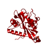
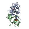
| Title | x-ray structure of cj1430 in the presence of GDP, a GDP-D-glycero-4-keto-D-lyxo-heptose-3,5-epimerase from campylobacter jejuni | |||||||||
 Components Components | Thymidine diphospho-4-keto-rhamnose 3,5-epimerase | |||||||||
 Keywords Keywords |  ISOMERASE / 3 / 5-epimerase / capsular polycassharide ISOMERASE / 3 / 5-epimerase / capsular polycassharide | |||||||||
| Function / homology |  Function and homology informationdTDP-4-dehydrorhamnose 3,5-epimerase / dTDP-4-dehydrorhamnose 3,5-epimerase activity / dTDP-rhamnose biosynthetic process / extracellular polysaccharide biosynthetic process / cytosol Function and homology informationdTDP-4-dehydrorhamnose 3,5-epimerase / dTDP-4-dehydrorhamnose 3,5-epimerase activity / dTDP-rhamnose biosynthetic process / extracellular polysaccharide biosynthetic process / cytosolSimilarity search - Function | |||||||||
| Biological species |  Campylobacter jejuni subsp. jejuni serotype O:2 (Campylobacter) Campylobacter jejuni subsp. jejuni serotype O:2 (Campylobacter) | |||||||||
| Method | X-RAY DIFFRACTION / MOLECULAR REPLACEMENT / Resolution: 2.1 Å | |||||||||
 Authors Authors | Girardi, N.M. / Thoden, J.B. / Raushel, F.M. / Holden, H.M. | |||||||||
| Funding support |  United States, 2items United States, 2items
| |||||||||
 Citation Citation | Journal: Biochemistry / Year: 2021 Title: Biosynthesis of d- glycero -l- gluco -Heptose in the Capsular Polysaccharides of Campylobacter jejuni . Authors: Huddleston, J.P. / Anderson, T.K. / Girardi, N.M. / Thoden, J.B. / Taylor, Z. / Holden, H.M. / Raushel, F.M. | |||||||||
| History |
|
- Structure visualization
| Structure viewer | Molecule: MolmilJmol/JSmol |
|---|
- Downloads & links
-Download
| PDBx/mmCIF format | 7m14.cif.gz | 244 KB | Display | PDBx/mmCIF format |
|---|---|---|---|---|
| PDB format | pdb7m14.ent.gz | 194.9 KB | Display | PDB format |
| PDBx/mmJSON format | 7m14.json.gz | Tree view | PDBx/mmJSON format | |
| Others |  Other downloads Other downloads |
-Validation report
| Arichive directory | https://data.pdbj.org/pub/pdb/validation_reports/m1/7m14ftp://data.pdbj.org/pub/pdb/validation_reports/m1/7m14 | HTTPS FTP |
|---|
-Related structure data
| Related structure data |  7m13C  7m15C  3rykS S: Starting model for refinement C: citing same article ( |
|---|---|
| Similar structure data |


-Links
 PDBj
PDBj- Assembly
| Deposited unit | 
| ||||||||
|---|---|---|---|---|---|---|---|---|---|
| 1 | 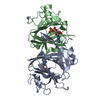
| ||||||||
| 2 | 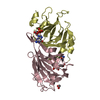
| ||||||||
| 3 | 
| ||||||||
| Unit cell |
| ||||||||
| Components on special symmetry positions |
|
-Components
| #1: Protein | Mass: 21365.320 Da / Num. of mol.: 6 / Mutation: K128A, E129A Source method: isolated from a genetically manipulated source Source: (gene. exp.) Campylobacter jejuni subsp. jejuni serotype O:2 (strain ATCC 700819 / NCTC 11168) (Campylobacter)Strain: ATCC 700819 / NCTC 11168 / Gene: rfbC, Cj1430c / Production host: Escherichia coli (E. coli)References: UniProt: Q0P8I4, dTDP-4-dehydrorhamnose 3,5-epimerase#2: Chemical | ChemComp-GDP / Guanosine diphosphate  Type: RNA linking / Mass: 443.201 Da / Num. of mol.: 6 / Source method: obtained synthetically / Formula: C10H15N5O11P2 / Comment: GDP, energy-carrying molecule*YM Type: RNA linking / Mass: 443.201 Da / Num. of mol.: 6 / Source method: obtained synthetically / Formula: C10H15N5O11P2 / Comment: GDP, energy-carrying molecule*YM#3: Chemical | ChemComp-NA / |   Mass: 22.990 Da / Num. of mol.: 1 / Source method: obtained synthetically / Formula: Na Mass: 22.990 Da / Num. of mol.: 1 / Source method: obtained synthetically / Formula: Na#4: Chemical | Ethylene glycol  Mass: 62.068 Da / Num. of mol.: 2 / Source method: obtained synthetically / Formula: C2H6O2 Mass: 62.068 Da / Num. of mol.: 2 / Source method: obtained synthetically / Formula: C2H6O2#5: Water | ChemComp-HOH / | Water Mass: 18.015 Da / Num. of mol.: 645 / Source method: isolated from a natural source / Formula: H2O Mass: 18.015 Da / Num. of mol.: 645 / Source method: isolated from a natural source / Formula: H2OHas ligand of interest | N | |
|---|
-Experimental details
-Experiment
| Experiment | Method: X-RAY DIFFRACTION / Number of used crystals: 1 |
|---|
- Sample preparation
| Crystal | Density Matthews: 2.4 Å3/Da / Density % sol: 48.84 % |
|---|---|
| Crystal grow | Temperature: 293 K / Method: vapor diffusion, hanging drop / pH: 7 / Details: 22-27% PEG-5000, 100 mM MOPS, 10 mM GDP |
-Data collection
| Diffraction | Mean temperature: 100 K / Serial crystal experiment: N |
|---|---|
| Diffraction source | Source: SEALED TUBE / Type: BRUKER D8 QUEST / Wavelength: 1.5418 Å |
| Detector | Type: Bruker PHOTON II / Detector: PIXEL / Date: Aug 29, 2020 |
| Radiation | Protocol: SINGLE WAVELENGTH / Monochromatic (M) / Laue (L): M / Scattering type: x-ray |
| Radiation wavelength | Wavelength: 1.5418 Å / Relative weight: 1 |
| Reflection | Resolution: 2.1→50 Å / Num. obs: 69127 / % possible obs: 97.3 % / Observed criterion σ(F): 0 / Observed criterion σ(I): 0 / Redundancy: 5.2 % / Rsym value: 0.088 / Net I/σ(I): 9.7 |
| Reflection shell | Resolution: 2.1→2.2 Å / Mean I/σ(I) obs: 2.4 / Num. unique obs: 8583 / Rsym value: 0.39 / % possible all: 93 |
- Processing
| Software |
| ||||||||||||||||||||||||||||||||||||||||||||||||||||||||||||
|---|---|---|---|---|---|---|---|---|---|---|---|---|---|---|---|---|---|---|---|---|---|---|---|---|---|---|---|---|---|---|---|---|---|---|---|---|---|---|---|---|---|---|---|---|---|---|---|---|---|---|---|---|---|---|---|---|---|---|---|---|---|
| Refinement | Method to determine structure: MOLECULAR REPLACEMENT Starting model: 3ryk Resolution: 2.1→27.83 Å / Cor.coef. Fo:Fc: 0.941 / Cor.coef. Fo:Fc free: 0.89 / SU B: 7.744 / SU ML: 0.19 / Cross valid method: THROUGHOUT / σ(F): 0 / ESU R: 0.258 / ESU R Free: 0.217 / Stereochemistry target values: MAXIMUM LIKELIHOOD Details: HYDROGENS HAVE BEEN ADDED IN THE RIDING POSITIONS U VALUES : REFINED INDIVIDUALLY
| ||||||||||||||||||||||||||||||||||||||||||||||||||||||||||||
| Solvent computation | Ion probe radii: 0.8 Å / Shrinkage radii: 0.8 Å / VDW probe radii: 1.2 Å / Solvent model: MASK | ||||||||||||||||||||||||||||||||||||||||||||||||||||||||||||
| Displacement parameters | Biso max: 90.69 Å2 / Biso mean: 26.749 Å2 / Biso min: 5.08 Å2
| ||||||||||||||||||||||||||||||||||||||||||||||||||||||||||||
| Refinement step | Cycle: final / Resolution: 2.1→27.83 Å
| ||||||||||||||||||||||||||||||||||||||||||||||||||||||||||||
| Refine LS restraints |
| ||||||||||||||||||||||||||||||||||||||||||||||||||||||||||||
| LS refinement shell | Resolution: 2.1→2.154 Å / Rfactor Rfree error: 0 / Total num. of bins used: 20
|
