 Movie
Movie Controller

[English] 日本語
 Yorodumi
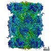
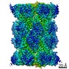
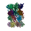
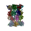
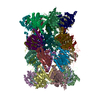
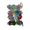
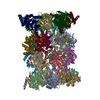
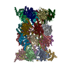
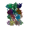
- PDB-7lxu: Structure of Plasmodium falciparum 20S proteasome with bound MPI-5 -
+ Open data

- Basic information
| Entry | Database: PDB / ID: 7lxu | |||||||||
|---|---|---|---|---|---|---|---|---|---|---|
| Title | Structure of Plasmodium falciparum 20S proteasome with bound MPI-5 | |||||||||
 Components Components |
| |||||||||
 Keywords Keywords |  HYDROLASE / proteasome / plasmodium falciparum / malaria / drug / bortezomib HYDROLASE / proteasome / plasmodium falciparum / malaria / drug / bortezomib | |||||||||
| Function / homology |  Function and homology information Function and homology informationCross-presentation of soluble exogenous antigens (endosomes) / : / Orc1 removal from chromatin / CDK-mediated phosphorylation and removal of Cdc6 / KEAP1-NFE2L2 pathway / UCH proteinases / Ub-specific processing proteases / Neddylation / Antigen processing: Ubiquitination & Proteasome degradation / Neutrophil degranulation ...Cross-presentation of soluble exogenous antigens (endosomes) / : / Orc1 removal from chromatin / CDK-mediated phosphorylation and removal of Cdc6 / KEAP1-NFE2L2 pathway / UCH proteinases / Ub-specific processing proteases / Neddylation / Antigen processing: Ubiquitination & Proteasome degradation / Neutrophil degranulation / proteasome core complex / proteasome endopeptidase complex / proteasome core complex, beta-subunit complex / proteasome core complex, alpha-subunit complex / threonine-type endopeptidase activity / proteasomal protein catabolic process / ubiquitin-dependent protein catabolic process / proteasome-mediated ubiquitin-dependent protein catabolic process / endopeptidase activity / hydrolase activity / nucleus / cytosol / cytoplasmSimilarity search - Function | |||||||||
| Biological species |  Plasmodium falciparum (malaria parasite P. falciparum) Plasmodium falciparum (malaria parasite P. falciparum) | |||||||||
| Method | ELECTRON MICROSCOPY / single particle reconstruction / cryo EM / Resolution: 3.1 Å | |||||||||
 Authors Authors | Metcalfe, R.D. / Morton, C.J. / Xie, S.C. / Liu, B. / Hanssen, E. / Leis, A.P. / Tilley, L. / Griffin, M.D.W. | |||||||||
| Funding support |  Australia, Australia,  United States, 2items United States, 2items
| |||||||||
 Citation Citation | Journal: Proc Natl Acad Sci U S A / Year: 2021 Title: Design of proteasome inhibitors with oral efficacy in vivo against and selectivity over the human proteasome. Authors: Stanley C Xie / Riley D Metcalfe / Hirotake Mizutani / Tanya Puhalovich / Eric Hanssen / Craig J Morton / Yawei Du / Con Dogovski / Shih-Chung Huang / Jeffrey Ciavarri / Paul Hales / Robert ...Authors: Stanley C Xie / Riley D Metcalfe / Hirotake Mizutani / Tanya Puhalovich / Eric Hanssen / Craig J Morton / Yawei Du / Con Dogovski / Shih-Chung Huang / Jeffrey Ciavarri / Paul Hales / Robert J Griffin / Lawrence H Cohen / Bei-Ching Chuang / Sergio Wittlin / Ioanna Deni / Tomas Yeo / Kurt E Ward / Daniel C Barry / Boyin Liu / David L Gillett / Benigno F Crespo-Fernandez / Sabine Ottilie / Nimisha Mittal / Alisje Churchyard / Daniel Ferguson / Anna Caroline C Aguiar / Rafael V C Guido / Jake Baum / Kirsten K Hanson / Elizabeth A Winzeler / Francisco-Javier Gamo / David A Fidock / Delphine Baud / Michael W Parker / Stephen Brand / Lawrence R Dick / Michael D W Griffin / Alexandra E Gould / Leann Tilley /     Abstract: The proteasome is a potential antimalarial drug target. We have identified a series of amino-amide boronates that are potent and specific inhibitors of the 20S proteasome (20S) β5 active site and ...The proteasome is a potential antimalarial drug target. We have identified a series of amino-amide boronates that are potent and specific inhibitors of the 20S proteasome (20S) β5 active site and that exhibit fast-acting antimalarial activity. They selectively inhibit the growth of compared with a human cell line and exhibit high potency against field isolates of and They have a low propensity for development of resistance and possess liver stage and transmission-blocking activity. Exemplar compounds, MPI-5 and MPI-13, show potent activity against infections in a SCID mouse model with an oral dosing regimen that is well tolerated. We show that MPI-5 binds more strongly to 20S than to human constitutive 20S (20Sc). Comparison of the cryo-electron microscopy (EM) structures of 20S and 20Sc in complex with MPI-5 and 20S in complex with the clinically used anti-cancer agent, bortezomib, reveal differences in binding modes that help to explain the selectivity. Together, this work provides insights into the 20S proteasome in , underpinning the design of potent and selective antimalarial proteasome inhibitors. | |||||||||
| History |
|
- Structure visualization
| Movie |
Movie viewer |
|---|---|
| Structure viewer | Molecule: MolmilJmol/JSmol |
- Downloads & links
-Download
| PDBx/mmCIF format | 7lxu.cif.gz | 1.1 MB | Display | PDBx/mmCIF format |
|---|---|---|---|---|
| PDB format | pdb7lxu.ent.gz | 919.7 KB | Display | PDB format |
| PDBx/mmJSON format | 7lxu.json.gz | Tree view | PDBx/mmJSON format | |
| Others |  Other downloads Other downloads |
-Validation report
| Arichive directory | https://data.pdbj.org/pub/pdb/validation_reports/lx/7lxuftp://data.pdbj.org/pub/pdb/validation_reports/lx/7lxu | HTTPS FTP |
|---|
-Related structure data
| Related structure data | 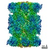 23575MC 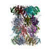 7lxtC 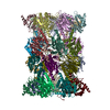 7lxvC M: map data used to model this data C: citing same article ( |
|---|---|
| Similar structure data |


-Links
 PDBj
PDBj


- Assembly
| Deposited unit | 
|
|---|---|
| 1 |
|
-Components
-20S proteasome alpha- ... , 7 types, 14 molecules AOBPCQDRESFTGU
| #1: Protein | Mass: 29531.656 Da / Num. of mol.: 2 / Source method: isolated from a natural source Source: (natural) Plasmodium falciparum (isolate 3D7) (eukaryote)Strain: isolate 3D7 References: UniProt: Q8IAR3, proteasome endopeptidase complex#2: Protein | Mass: 26556.391 Da / Num. of mol.: 2 / Source method: isolated from a natural source Source: (natural) Plasmodium falciparum (isolate 3D7) (eukaryote)Strain: isolate 3D7 References: UniProt: C6KST3, proteasome endopeptidase complex#3: Protein | Mass: 27977.664 Da / Num. of mol.: 2 / Source method: isolated from a natural source Source: (natural) Plasmodium falciparum (isolate 3D7) (eukaryote)Strain: isolate 3D7 References: UniProt: Q8IDG3, proteasome endopeptidase complex#4: Protein | Mass: 27263.285 Da / Num. of mol.: 2 / Source method: isolated from a natural source Source: (natural) Plasmodium falciparum (isolate 3D7) (eukaryote)Strain: isolate 3D7 References: UniProt: Q8IDG2, proteasome endopeptidase complex#5: Protein | Mass: 28417.367 Da / Num. of mol.: 2 / Source method: isolated from a natural source Source: (natural) Plasmodium falciparum (isolate 3D7) (eukaryote)Strain: isolate 3D7 References: UniProt: Q8IBI3, proteasome endopeptidase complex#6: Protein | Mass: 28871.697 Da / Num. of mol.: 2 / Source method: isolated from a natural source Source: (natural) Plasmodium falciparum (isolate 3D7) (eukaryote)Strain: isolate 3D7 References: UniProt: Q8IK90, proteasome endopeptidase complex#7: Protein | Mass: 29324.295 Da / Num. of mol.: 2 / Source method: isolated from a natural source Source: (natural) Plasmodium falciparum (isolate 3D7) (eukaryote)Strain: isolate 3D7 References: UniProt: O77396, proteasome endopeptidase complex |
|---|
-20S proteasome beta- ... , 7 types, 14 molecules HVIWJXKYLZMaNb
| #8: Protein | Mass: 29143.936 Da / Num. of mol.: 2 / Source method: isolated from a natural source Source: (natural) Plasmodium falciparum (isolate 3D7) (eukaryote)Strain: isolate 3D7 References: UniProt: Q8I0U7, proteasome endopeptidase complex#9: Protein | Mass: 25104.885 Da / Num. of mol.: 2 / Source method: isolated from a natural source Source: (natural) Plasmodium falciparum (isolate 3D7) (eukaryote)Strain: isolate 3D7 References: UniProt: Q8I6T3, proteasome endopeptidase complex#10: Protein | Mass: 24533.131 Da / Num. of mol.: 2 / Source method: isolated from a natural source Source: (natural) Plasmodium falciparum (isolate 3D7) (eukaryote)Strain: isolate 3D7 References: UniProt: Q8I261, proteasome endopeptidase complex#11: Protein | Mass: 22889.105 Da / Num. of mol.: 2 / Source method: isolated from a natural source Source: (natural) Plasmodium falciparum (isolate 3D7) (eukaryote)Strain: isolate 3D7 References: UniProt: Q8IKC9, proteasome endopeptidase complex#12: Protein | Mass: 23620.646 Da / Num. of mol.: 2 / Source method: isolated from a natural source Source: (natural) Plasmodium falciparum (isolate 3D7) (eukaryote)Strain: isolate 3D7 References: UniProt: Q8IJT1, proteasome endopeptidase complex#13: Protein | Mass: 27301.203 Da / Num. of mol.: 2 / Source method: isolated from a natural source Source: (natural) Plasmodium falciparum (isolate 3D7) (eukaryote)Strain: isolate 3D7 References: UniProt: A0A5K1K7U1, proteasome endopeptidase complex#14: Protein | Mass: 30909.893 Da / Num. of mol.: 2 / Source method: isolated from a natural source Source: (natural) Plasmodium falciparum (isolate 3D7) (eukaryote)Strain: isolate 3D7 References: UniProt: Q7K6A9, proteasome endopeptidase complex |
|---|
-Non-polymers , 1 types, 2 molecules 
| #15: Chemical |  Mass: 352.235 Da / Num. of mol.: 2 / Source method: obtained synthetically / Formula: C20H25BN2O3 / Feature type: SUBJECT OF INVESTIGATION Mass: 352.235 Da / Num. of mol.: 2 / Source method: obtained synthetically / Formula: C20H25BN2O3 / Feature type: SUBJECT OF INVESTIGATION |
|---|
-Details
| Has ligand of interest | Y |
|---|
-Experimental details
-Experiment
| Experiment | Method: ELECTRON MICROSCOPY |
|---|---|
| EM experiment | Aggregation state: PARTICLE / 3D reconstruction method: single particle reconstruction |
- Sample preparation
| Component | Name: Plasmodium falciparum 20S proteasome complexed with ML052 Type: COMPLEX / Entity ID: #1-#11, #13-#14 / Source: NATURAL |
|---|---|
| Molecular weight | Value: 0.7 MDa / Experimental value: NO |
| Source (natural) | Organism: Plasmodium falciparum (malaria parasite P. falciparum) |
| Buffer solution | pH: 7.4 |
| Specimen | Embedding applied: NO / Shadowing applied: NO / Staining applied: NO / Vitrification applied: YES |
| Specimen support | Grid material: GOLD / Grid mesh size: 300 divisions/in. / Grid type: Quantifoil R1.2/1.3 |
| Vitrification | Instrument: FEI VITROBOT MARK III / Cryogen name: ETHANE / Humidity: 100 % / Chamber temperature: 295 K |
- Electron microscopy imaging
| Experimental equipment |  Model: Talos Arctica / Image courtesy: FEI Company |
|---|---|
| Microscopy | Model: FEI TALOS ARCTICA |
| Electron gun | Electron source: FIELD EMISSION GUN / Accelerating voltage: 200 kV / Illumination mode: FLOOD BEAM |
| Electron lens | Mode: BRIGHT FIELDBright-field microscopy / C2 aperture diameter: 50 µm |
| Image recording | Electron dose: 50 e/Å2 / Detector mode: SUPER-RESOLUTION / Film or detector model: GATAN K2 SUMMIT (4k x 4k) |
- Processing
| Software | Name: PHENIX / Version: 1.18.2_3874: / Classification: refinement | ||||||||||||||||||||||||
|---|---|---|---|---|---|---|---|---|---|---|---|---|---|---|---|---|---|---|---|---|---|---|---|---|---|
| EM software |
| ||||||||||||||||||||||||
| CTF correction | Type: PHASE FLIPPING AND AMPLITUDE CORRECTION | ||||||||||||||||||||||||
| Symmetry | Point symmetry: C2 (2 fold cyclic) | ||||||||||||||||||||||||
| 3D reconstruction | Resolution: 3.1 Å / Resolution method: FSC 0.143 CUT-OFF / Num. of particles: 38738 / Symmetry type: POINT | ||||||||||||||||||||||||
| Refine LS restraints |
|
