 Movie
Movie Controller

[English] 日本語
 Yorodumi
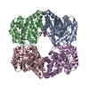
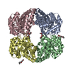
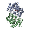
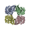
- PDB-7lvl: Dihydrodipicolinate synthase bound with allosteric inhibitor (S)-... -
+ Open data

- Basic information
| Entry | Database: PDB / ID: 7lvl | ||||||
|---|---|---|---|---|---|---|---|
| Title | Dihydrodipicolinate synthase bound with allosteric inhibitor (S)-lysine from Candidatus Liberibacter solanacearum | ||||||
 Components Components | 4-hydroxy-tetrahydrodipicolinate synthase Dihydrodipicolinate synthase Dihydrodipicolinate synthase | ||||||
 Keywords Keywords | LYASE / TIM barrel | ||||||
| Function / homology |  Function and homology information4-hydroxy-tetrahydrodipicolinate synthase / 4-hydroxy-tetrahydrodipicolinate synthase activity / diaminopimelate biosynthetic process / lysine biosynthetic process via diaminopimelate / cytoplasm Function and homology information4-hydroxy-tetrahydrodipicolinate synthase / 4-hydroxy-tetrahydrodipicolinate synthase activity / diaminopimelate biosynthetic process / lysine biosynthetic process via diaminopimelate / cytoplasmSimilarity search - Function | ||||||
| Biological species |  Candidatus Liberibacter solanacearum (bacteria) Candidatus Liberibacter solanacearum (bacteria) | ||||||
| Method | X-RAY DIFFRACTION / SYNCHROTRON / MOLECULAR REPLACEMENT / Resolution: 2.01 Å | ||||||
 Authors Authors | Gilkes, J.M. / Frampton, R.A. / Board, A.J. / Sheen, C.R. / Smith, G.R. / Dobson, R.C.J.D. | ||||||
 Citation Citation | Journal: To Be Published Title: Dihydrodipicolinate synthase bound with allosteric inhibitor (S)-lysine from Candidatus Liberibacter solanacearum Authors: Gilkes, J.M. / Frampton, R.A. / Board, A.J. / Sheen, C.R. / Smith, G.R. / Dobson, R.C.J.D. | ||||||
| History |
|
- Structure visualization
| Structure viewer | Molecule: MolmilJmol/JSmol |
|---|
- Downloads & links
-Download
| PDBx/mmCIF format | 7lvl.cif.gz | 688.7 KB | Display | PDBx/mmCIF format |
|---|---|---|---|---|
| PDB format | pdb7lvl.ent.gz | 573.6 KB | Display | PDB format |
| PDBx/mmJSON format | 7lvl.json.gz | Tree view | PDBx/mmJSON format | |
| Others |  Other downloads Other downloads |
-Validation report
| Arichive directory | https://data.pdbj.org/pub/pdb/validation_reports/lv/7lvlftp://data.pdbj.org/pub/pdb/validation_reports/lv/7lvl | HTTPS FTP |
|---|
-Related structure data
| Related structure data |  7loyS S: Starting model for refinement |
|---|---|
| Similar structure data |


-Links
 PDBj
PDBj- Assembly
| Deposited unit | 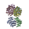
| ||||||||
|---|---|---|---|---|---|---|---|---|---|
| 1 | 
| ||||||||
| 2 | 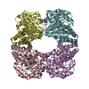
| ||||||||
| Unit cell |
| ||||||||
| Components on special symmetry positions |
|
-Components
| #1: Protein | Dihydrodipicolinate synthase / HTPA synthase Mass: 32695.988 Da / Num. of mol.: 6 Source method: isolated from a genetically manipulated source Source: (gene. exp.) Candidatus Liberibacter solanacearum (bacteria)Gene: dapA, DJ66_0589 / Production host: Escherichia coli BL21 (bacteria)References: UniProt: A0A0F4VK59, 4-hydroxy-tetrahydrodipicolinate synthase#2: Chemical | ChemComp-LYS / Lysine  Type: L-peptide linking / Mass: 147.195 Da / Num. of mol.: 6 / Source method: obtained synthetically / Formula: C6H15N2O2 / Feature type: SUBJECT OF INVESTIGATION Type: L-peptide linking / Mass: 147.195 Da / Num. of mol.: 6 / Source method: obtained synthetically / Formula: C6H15N2O2 / Feature type: SUBJECT OF INVESTIGATION#3: Water | ChemComp-HOH / | Water Mass: 18.015 Da / Num. of mol.: 876 / Source method: isolated from a natural source / Formula: H2O Mass: 18.015 Da / Num. of mol.: 876 / Source method: isolated from a natural source / Formula: H2OHas ligand of interest | Y | |
|---|
-Experimental details
-Experiment
| Experiment | Method: X-RAY DIFFRACTION / Number of used crystals: 1 |
|---|
- Sample preparation
| Crystal | Density Matthews: 2.64 Å3/Da / Density % sol: 54.61 % |
|---|---|
| Crystal grow | Temperature: 293.15 K / Method: vapor diffusion, sitting drop / pH: 5.5 / Details: 20% w/v PEG3000, 0.1 M sodium citrate, pH 5.5 |
-Data collection
| Diffraction | Mean temperature: 210 K / Serial crystal experiment: N |
|---|---|
| Diffraction source | Source: SYNCHROTRON / Site: Australian Synchrotron  / Beamline: MX2 / Wavelength: 0.95372 Å / Beamline: MX2 / Wavelength: 0.95372 Å |
| Detector | Type: DECTRIS EIGER X 16M / Detector: PIXEL / Date: Dec 7, 2018 |
| Radiation | Protocol: SINGLE WAVELENGTH / Monochromatic (M) / Laue (L): M / Scattering type: x-ray |
| Radiation wavelength | Wavelength: 0.95372 Å / Relative weight: 1 |
| Reflection | Resolution: 2.01→46.24 Å / Num. obs: 135620 / % possible obs: 99.92 % / Redundancy: 4 % / Biso Wilson estimate: 31.38 Å2 / CC1/2: 0.954 / Net I/σ(I): 13.45 |
| Reflection shell | Resolution: 2.01→2.082 Å / Num. unique obs: 13469 / CC1/2: 0.802 |
- Processing
| Software |
| ||||||||||||||||||||||||||||||||||||||||||||||||||||||||||||||||||||||||||||||||||||||||||||||||||||||||||||||||||||||||||||||||||||||||||||||||||||||||||||||||||||||||||||||||||||||||||||||||||||||||||||||||||
|---|---|---|---|---|---|---|---|---|---|---|---|---|---|---|---|---|---|---|---|---|---|---|---|---|---|---|---|---|---|---|---|---|---|---|---|---|---|---|---|---|---|---|---|---|---|---|---|---|---|---|---|---|---|---|---|---|---|---|---|---|---|---|---|---|---|---|---|---|---|---|---|---|---|---|---|---|---|---|---|---|---|---|---|---|---|---|---|---|---|---|---|---|---|---|---|---|---|---|---|---|---|---|---|---|---|---|---|---|---|---|---|---|---|---|---|---|---|---|---|---|---|---|---|---|---|---|---|---|---|---|---|---|---|---|---|---|---|---|---|---|---|---|---|---|---|---|---|---|---|---|---|---|---|---|---|---|---|---|---|---|---|---|---|---|---|---|---|---|---|---|---|---|---|---|---|---|---|---|---|---|---|---|---|---|---|---|---|---|---|---|---|---|---|---|---|---|---|---|---|---|---|---|---|---|---|---|---|---|---|---|---|
| Refinement | Method to determine structure: MOLECULAR REPLACEMENT Starting model: PDB entry 7LOY Resolution: 2.01→46.202 Å / SU ML: 0.2 / Cross valid method: THROUGHOUT / σ(F): 1.94 / Phase error: 19.74 / Stereochemistry target values: MLHL
| ||||||||||||||||||||||||||||||||||||||||||||||||||||||||||||||||||||||||||||||||||||||||||||||||||||||||||||||||||||||||||||||||||||||||||||||||||||||||||||||||||||||||||||||||||||||||||||||||||||||||||||||||||
| Solvent computation | Shrinkage radii: 0.9 Å / VDW probe radii: 1.11 Å / Solvent model: FLAT BULK SOLVENT MODEL | ||||||||||||||||||||||||||||||||||||||||||||||||||||||||||||||||||||||||||||||||||||||||||||||||||||||||||||||||||||||||||||||||||||||||||||||||||||||||||||||||||||||||||||||||||||||||||||||||||||||||||||||||||
| Refinement step | Cycle: LAST / Resolution: 2.01→46.202 Å
| ||||||||||||||||||||||||||||||||||||||||||||||||||||||||||||||||||||||||||||||||||||||||||||||||||||||||||||||||||||||||||||||||||||||||||||||||||||||||||||||||||||||||||||||||||||||||||||||||||||||||||||||||||
| Refine LS restraints |
| ||||||||||||||||||||||||||||||||||||||||||||||||||||||||||||||||||||||||||||||||||||||||||||||||||||||||||||||||||||||||||||||||||||||||||||||||||||||||||||||||||||||||||||||||||||||||||||||||||||||||||||||||||
| LS refinement shell |
| ||||||||||||||||||||||||||||||||||||||||||||||||||||||||||||||||||||||||||||||||||||||||||||||||||||||||||||||||||||||||||||||||||||||||||||||||||||||||||||||||||||||||||||||||||||||||||||||||||||||||||||||||||
| Refinement TLS params. | Method: refined / Origin x: 17.2875 Å / Origin y: -2.2409 Å / Origin z: 42.7079 Å
| ||||||||||||||||||||||||||||||||||||||||||||||||||||||||||||||||||||||||||||||||||||||||||||||||||||||||||||||||||||||||||||||||||||||||||||||||||||||||||||||||||||||||||||||||||||||||||||||||||||||||||||||||||
| Refinement TLS group | Selection details: all |
