 Movie
Movie Controller


+ Open data

- Basic information
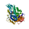
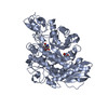
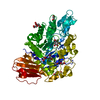
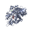
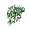
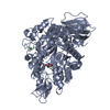
| Entry | Database: PDB / ID: 7lv6 | ||||||
|---|---|---|---|---|---|---|---|
| Title | The structure of MalL mutant enzyme S536R from Bacillus subtilis | ||||||
 Components Components | Oligo-1,6-glucosidase 1 Sucrase-isomaltase Sucrase-isomaltase | ||||||
 Keywords Keywords | HYDROLASE / TIM barrel / glycoside hydrolase / enzyme design / Rosetta | ||||||
| Function / homology |  Function and homology informationoligo-1,6-glucosidase / oligo-1,6-glucosidase activity / oligosaccharide catabolic process / alpha-amylase activity / metal ion binding / cytoplasm Function and homology informationoligo-1,6-glucosidase / oligo-1,6-glucosidase activity / oligosaccharide catabolic process / alpha-amylase activity / metal ion binding / cytoplasmSimilarity search - Function | ||||||
| Biological species |  Bacillus subtilis (bacteria) Bacillus subtilis (bacteria) | ||||||
| Method | X-RAY DIFFRACTION / SYNCHROTRON / MOLECULAR REPLACEMENT / molecular replacement / Resolution: 1.1 Å | ||||||
 Authors Authors | Hamill, C.J. / Prentice, E.J. / Bahl, C.D. / Truebridge, I.S. / Arcus, V.L. | ||||||
| Funding support |  New Zealand, 1items New Zealand, 1items
| ||||||
 Citation Citation | Journal: To Be Published Title: Urea binding to guide rational design of mutations that influence enzyme dynamics Authors: Hamill, C.J. / Arcus, V.L. / Prentice, E.J. / Bahl, C. / Truebridge, I. | ||||||
| History |
|
- Structure visualization
| Structure viewer | Molecule: MolmilJmol/JSmol |
|---|
- Downloads & links
-Download
| PDBx/mmCIF format | 7lv6.cif.gz | 370.2 KB | Display | PDBx/mmCIF format |
|---|---|---|---|---|
| PDB format | pdb7lv6.ent.gz | 301.2 KB | Display | PDB format |
| PDBx/mmJSON format | 7lv6.json.gz | Tree view | PDBx/mmJSON format | |
| Others |  Other downloads Other downloads |
-Validation report
| Arichive directory | https://data.pdbj.org/pub/pdb/validation_reports/lv/7lv6ftp://data.pdbj.org/pub/pdb/validation_reports/lv/7lv6 | HTTPS FTP |
|---|
-Related structure data
| Related structure data | 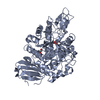 4m56S S: Starting model for refinement |
|---|---|
| Similar structure data |


-Links
 PDBj
PDBj

- Assembly
| Deposited unit | 
| ||||||||
|---|---|---|---|---|---|---|---|---|---|
| 1 |
| ||||||||
| Unit cell |
|
-Components
| #1: Protein | Sucrase-isomaltase / Dextrin 6-alpha-D-glucanohydrolase / Oligosaccharide alpha-1 / 6-glucosidase 1 / Sucrase-isomaltase ...Dextrin 6-alpha-D-glucanohydrolase / Oligosaccharide alpha-1 / 6-glucosidase 1 / Sucrase-isomaltase 1 / Isomaltase 1 Mass: 69305.555 Da / Num. of mol.: 1 / Mutation: S536R Source method: isolated from a genetically manipulated source Source: (gene. exp.) Bacillus subtilis (strain 168) (bacteria)Strain: 168 / Gene: malL, yvdL, BSU34560 / Plasmid: pPROEX-Htb / Production host: Escherichia coli (E. coli) / Strain (production host): DH5a / References: UniProt: O06994, oligo-1,6-glucosidase |
|---|---|
| #2: Chemical | ChemComp-TRS / Tris  Mass: 122.143 Da / Num. of mol.: 1 / Source method: obtained synthetically / Formula: C4H12NO3 / Comment: pH buffer*YM Mass: 122.143 Da / Num. of mol.: 1 / Source method: obtained synthetically / Formula: C4H12NO3 / Comment: pH buffer*YM |
| #3: Chemical | ChemComp-GOL / Glycerol  Mass: 92.094 Da / Num. of mol.: 1 / Source method: obtained synthetically / Formula: C3H8O3 Mass: 92.094 Da / Num. of mol.: 1 / Source method: obtained synthetically / Formula: C3H8O3 |
| #4: Chemical | ChemComp-CA /   Mass: 40.078 Da / Num. of mol.: 1 / Source method: obtained synthetically / Formula: Ca Mass: 40.078 Da / Num. of mol.: 1 / Source method: obtained synthetically / Formula: Ca |
| #5: Water | ChemComp-HOH / Water Mass: 18.015 Da / Num. of mol.: 785 / Source method: isolated from a natural source / Formula: H2O Mass: 18.015 Da / Num. of mol.: 785 / Source method: isolated from a natural source / Formula: H2O |
| Has ligand of interest | N |
-Experimental details
-Experiment
| Experiment | Method: X-RAY DIFFRACTION / Number of used crystals: 1 |
|---|
- Sample preparation
| Crystal | Density Matthews: 2.12 Å3/Da / Density % sol: 41.95 % |
|---|---|
| Crystal grow | Temperature: 291 K / Method: vapor diffusion, hanging drop / pH: 8 Details: 0.1 M Tris, pH 8.0, 0.2 M ammonium acetate, 18% w/v PEG10000 |
-Data collection
| Diffraction | Mean temperature: 100 K / Serial crystal experiment: N | |||||||||||||||||||||
|---|---|---|---|---|---|---|---|---|---|---|---|---|---|---|---|---|---|---|---|---|---|---|
| Diffraction source | Source: SYNCHROTRON / Site: Australian Synchrotron  / Beamline: MX2 / Wavelength: 0.953735 Å / Beamline: MX2 / Wavelength: 0.953735 Å | |||||||||||||||||||||
| Detector | Type: DECTRIS EIGER X 16M / Detector: PIXEL / Date: May 1, 2019 | |||||||||||||||||||||
| Radiation | Protocol: SINGLE WAVELENGTH / Monochromatic (M) / Laue (L): M / Scattering type: x-ray | |||||||||||||||||||||
| Radiation wavelength | Wavelength: 0.953735 Å / Relative weight: 1 | |||||||||||||||||||||
| Reflection | Resolution: 1.1→44.85 Å / Num. obs: 208774 / % possible obs: 94.2 % / Redundancy: 10.9 % / CC1/2: 0.998 / Rmerge(I) obs: 0.107 / Rpim(I) all: 0.031 / Rrim(I) all: 0.112 / Net I/σ(I): 12.7 / Num. measured all: 2270037 / Scaling rejects: 1914 | |||||||||||||||||||||
| Reflection shell | Diffraction-ID: 1 / Resolution: 1.1→1.12 Å
|
-Phasing
| Phasing | Method: molecular replacement | ||||||
|---|---|---|---|---|---|---|---|
| Phasing MR | R rigid body: 0.36
|
- Processing
| Software |
| ||||||||||||||||||||||||||||||||||||||||||||||||||||||||||||||||||||||||||||||||||||||||||||||||||||||||||||||||||||||||||||||||||||||||||||||||||||||||||||||||||||||||||||||||||||||||||
|---|---|---|---|---|---|---|---|---|---|---|---|---|---|---|---|---|---|---|---|---|---|---|---|---|---|---|---|---|---|---|---|---|---|---|---|---|---|---|---|---|---|---|---|---|---|---|---|---|---|---|---|---|---|---|---|---|---|---|---|---|---|---|---|---|---|---|---|---|---|---|---|---|---|---|---|---|---|---|---|---|---|---|---|---|---|---|---|---|---|---|---|---|---|---|---|---|---|---|---|---|---|---|---|---|---|---|---|---|---|---|---|---|---|---|---|---|---|---|---|---|---|---|---|---|---|---|---|---|---|---|---|---|---|---|---|---|---|---|---|---|---|---|---|---|---|---|---|---|---|---|---|---|---|---|---|---|---|---|---|---|---|---|---|---|---|---|---|---|---|---|---|---|---|---|---|---|---|---|---|---|---|---|---|---|---|---|---|
| Refinement | Method to determine structure: MOLECULAR REPLACEMENT Starting model: PDB entry 4M56 Resolution: 1.1→33.54 Å / SU ML: 0.08 / Cross valid method: THROUGHOUT / σ(F): 1.36 / Phase error: 12.95 / Stereochemistry target values: ML
| ||||||||||||||||||||||||||||||||||||||||||||||||||||||||||||||||||||||||||||||||||||||||||||||||||||||||||||||||||||||||||||||||||||||||||||||||||||||||||||||||||||||||||||||||||||||||||
| Solvent computation | Shrinkage radii: 0.9 Å / VDW probe radii: 1.11 Å / Solvent model: FLAT BULK SOLVENT MODEL | ||||||||||||||||||||||||||||||||||||||||||||||||||||||||||||||||||||||||||||||||||||||||||||||||||||||||||||||||||||||||||||||||||||||||||||||||||||||||||||||||||||||||||||||||||||||||||
| Displacement parameters | Biso max: 69.07 Å2 / Biso mean: 16.1338 Å2 / Biso min: 7.2 Å2 | ||||||||||||||||||||||||||||||||||||||||||||||||||||||||||||||||||||||||||||||||||||||||||||||||||||||||||||||||||||||||||||||||||||||||||||||||||||||||||||||||||||||||||||||||||||||||||
| Refinement step | Cycle: final / Resolution: 1.1→33.54 Å
| ||||||||||||||||||||||||||||||||||||||||||||||||||||||||||||||||||||||||||||||||||||||||||||||||||||||||||||||||||||||||||||||||||||||||||||||||||||||||||||||||||||||||||||||||||||||||||
| LS refinement shell | Refine-ID: X-RAY DIFFRACTION / Rfactor Rfree error: 0
|
