 Movie
Movie Controller


+ Open data

- Basic information
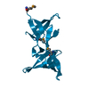
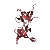
| Entry | Database: PDB / ID: 7liq | ||||||||||||
|---|---|---|---|---|---|---|---|---|---|---|---|---|---|
| Title | X-ray structure of SPOP MATH domain (S119A) | ||||||||||||
 Components Components | Speckle-type POZ protein | ||||||||||||
 Keywords Keywords |  PROTEIN BINDING / SPOP / 53BP1 / DNA damage response / Homologous recombination / Ubiquitin ligase PROTEIN BINDING / SPOP / 53BP1 / DNA damage response / Homologous recombination / Ubiquitin ligase | ||||||||||||
| Function / homology |  Function and homology informationregulation of proteolysis / Cul3-RING ubiquitin ligase complex / molecular function inhibitor activity / localization / Hedgehog 'on' state / protein polyubiquitination / proteasome-mediated ubiquitin-dependent protein catabolic process / nuclear speck / ubiquitin protein ligase binding / nucleoplasm ...regulation of proteolysis / Cul3-RING ubiquitin ligase complex / molecular function inhibitor activity / localization / Hedgehog 'on' state / protein polyubiquitination / proteasome-mediated ubiquitin-dependent protein catabolic process / nuclear speck / ubiquitin protein ligase binding / nucleoplasm / nucleus / cytoplasm Function and homology informationregulation of proteolysis / Cul3-RING ubiquitin ligase complex / molecular function inhibitor activity / localization / Hedgehog 'on' state / protein polyubiquitination / proteasome-mediated ubiquitin-dependent protein catabolic process / nuclear speck / ubiquitin protein ligase binding / nucleoplasm ...regulation of proteolysis / Cul3-RING ubiquitin ligase complex / molecular function inhibitor activity / localization / Hedgehog 'on' state / protein polyubiquitination / proteasome-mediated ubiquitin-dependent protein catabolic process / nuclear speck / ubiquitin protein ligase binding / nucleoplasm / nucleus / cytoplasmSimilarity search - Function | ||||||||||||
| Biological species |  Homo sapiens (human) Homo sapiens (human) | ||||||||||||
| Method | X-RAY DIFFRACTION / SYNCHROTRON / MOLECULAR REPLACEMENT / Resolution: 1.98 Å | ||||||||||||
 Authors Authors | Botuyan, M.V. / Cui, G. / Mer, G. | ||||||||||||
| Funding support |  United States, 3items United States, 3items
| ||||||||||||
 Citation Citation | Journal: Sci Adv / Year: 2021 Title: ATM-phosphorylated SPOP contributes to 53BP1 exclusion from chromatin during DNA replication. Authors: Wang, D. / Ma, J. / Botuyan, M.V. / Cui, G. / Yan, Y. / Ding, D. / Zhou, Y. / Krueger, E.W. / Pei, J. / Wu, X. / Wang, L. / Pei, H. / McNiven, M.A. / Ye, D. / Mer, G. / Huang, H. | ||||||||||||
| History |
|
- Structure visualization
| Structure viewer | Molecule: MolmilJmol/JSmol |
|---|
- Downloads & links
-Download
| PDBx/mmCIF format | 7liq.cif.gz | 84.4 KB | Display | PDBx/mmCIF format |
|---|---|---|---|---|
| PDB format | pdb7liq.ent.gz | 55 KB | Display | PDB format |
| PDBx/mmJSON format | 7liq.json.gz | Tree view | PDBx/mmJSON format | |
| Others |  Other downloads Other downloads |
-Validation report
| Arichive directory | https://data.pdbj.org/pub/pdb/validation_reports/li/7liqftp://data.pdbj.org/pub/pdb/validation_reports/li/7liq | HTTPS FTP |
|---|
-Related structure data
| Related structure data |  7linSC  7lioC 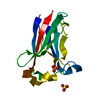 7lipC S: Starting model for refinement C: citing same article ( |
|---|---|
| Similar structure data |


-Links
 PDBj
PDBj


- Assembly
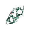
| Deposited unit | 
| ||||||||||||
|---|---|---|---|---|---|---|---|---|---|---|---|---|---|
| 1 |
| ||||||||||||
| Unit cell |
| ||||||||||||
| Components on special symmetry positions |
|
-Components
| #1: Protein | Mass: 16476.986 Da / Num. of mol.: 1 / Fragment: MATH domain / Mutation: S119A Source method: isolated from a genetically manipulated source Source: (gene. exp.) Homo sapiens (human) / Gene: SPOP / Production host:  Escherichia coli (E. coli) / References: UniProt: O43791 Escherichia coli (E. coli) / References: UniProt: O43791 |
|---|---|
| #2: Water | ChemComp-HOH / Water Mass: 18.015 Da / Num. of mol.: 64 / Source method: isolated from a natural source / Formula: H2O Mass: 18.015 Da / Num. of mol.: 64 / Source method: isolated from a natural source / Formula: H2O |
-Experimental details
-Experiment
| Experiment | Method: X-RAY DIFFRACTION / Number of used crystals: 1 |
|---|
- Sample preparation
| Crystal | Density Matthews: 2.79 Å3/Da / Density % sol: 55.94 % |
|---|---|
| Crystal grow | Temperature: 295 K / Method: vapor diffusion, hanging drop Details: Crystals were grown by the hanging drop method, mixing 2 ul of the SPOP MATH S119A at 20 mg/ml in 20 mM Tris-HCl, pH 7.6, 150 mM NaCl, 5 mM DTT and 2 ul of the reservoir solution for the ...Details: Crystals were grown by the hanging drop method, mixing 2 ul of the SPOP MATH S119A at 20 mg/ml in 20 mM Tris-HCl, pH 7.6, 150 mM NaCl, 5 mM DTT and 2 ul of the reservoir solution for the drop. The reservoir solution was 0.2 M (NH4)2SO4, 0.1 M BIS-Tris, pH 6.5, 25% (w/v) PEG 3,350). |
-Data collection
| Diffraction | Mean temperature: 100 K / Serial crystal experiment: N |
|---|---|
| Diffraction source | Source: SYNCHROTRON / Site: APS / Beamline: 19-ID / Wavelength: 0.9793 Å |
| Detector | Type: ADSC QUANTUM 315 / Detector: CCD / Date: Jul 25, 2019 |
| Radiation | Protocol: SINGLE WAVELENGTH / Monochromatic (M) / Laue (L): M / Scattering type: x-ray |
| Radiation wavelength | Wavelength: 0.9793 Å / Relative weight: 1 |
| Reflection | Resolution: 1.98→41.65 Å / Num. obs: 13677 / % possible obs: 99.98 % / Redundancy: 36.8 % / Biso Wilson estimate: 41.19 Å2 / CC1/2: 1 / CC star: 1 / Rmerge(I) obs: 0.067 / Rpim(I) all: 0.011 / Rrim(I) all: 0.068 / Net I/σ(I): 29.41 |
| Reflection shell | Resolution: 1.98→2.05 Å / Rmerge(I) obs: 0.998 / Num. unique obs: 1321 / CC1/2: 0.898 / CC star: 0.973 / Rpim(I) all: 0.182 / Rrim(I) all: 1.015 |
- Processing
| Software |
| |||||||||||||||||||||||||||||||||||||||||||||||||||||||||||||||||||||||||||||
|---|---|---|---|---|---|---|---|---|---|---|---|---|---|---|---|---|---|---|---|---|---|---|---|---|---|---|---|---|---|---|---|---|---|---|---|---|---|---|---|---|---|---|---|---|---|---|---|---|---|---|---|---|---|---|---|---|---|---|---|---|---|---|---|---|---|---|---|---|---|---|---|---|---|---|---|---|---|---|
| Refinement | Method to determine structure: MOLECULAR REPLACEMENT Starting model: 7LIN Resolution: 1.98→41.65 Å / SU ML: 0.2437 / Cross valid method: FREE R-VALUE / σ(F): 1.36 / Phase error: 21.6575 Stereochemistry target values: GeoStd + Monomer Library + CDL v1.2
| |||||||||||||||||||||||||||||||||||||||||||||||||||||||||||||||||||||||||||||
| Solvent computation | Shrinkage radii: 0.9 Å / VDW probe radii: 1.11 Å / Solvent model: FLAT BULK SOLVENT MODEL | |||||||||||||||||||||||||||||||||||||||||||||||||||||||||||||||||||||||||||||
| Displacement parameters | Biso mean: 55.36 Å2 | |||||||||||||||||||||||||||||||||||||||||||||||||||||||||||||||||||||||||||||
| Refinement step | Cycle: LAST / Resolution: 1.98→41.65 Å
| |||||||||||||||||||||||||||||||||||||||||||||||||||||||||||||||||||||||||||||
| Refine LS restraints |
| |||||||||||||||||||||||||||||||||||||||||||||||||||||||||||||||||||||||||||||
| LS refinement shell |
| |||||||||||||||||||||||||||||||||||||||||||||||||||||||||||||||||||||||||||||
| Refinement TLS params. | Method: refined / Refine-ID: X-RAY DIFFRACTION
| |||||||||||||||||||||||||||||||||||||||||||||||||||||||||||||||||||||||||||||
| Refinement TLS group |
|
