 Movie
Movie Controller

[English] 日本語
 Yorodumi
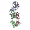
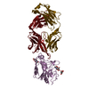
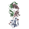
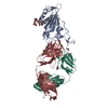
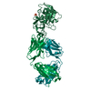
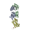
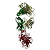
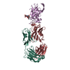
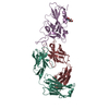
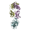
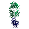
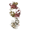
- PDB-7lcv: Factor H enhancing human antibody fragment (Fab) to meningococcal... -
+ Open data

- Basic information
| Entry | Database: PDB / ID: 7lcv | ||||||
|---|---|---|---|---|---|---|---|
| Title | Factor H enhancing human antibody fragment (Fab) to meningococcal Factor H binding protein | ||||||
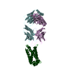
 Components Components |
| ||||||
 Keywords Keywords |  IMMUNE SYSTEM / human / antibody / Fab / meningococcal vaccine / Factor H binding protein / complex IMMUNE SYSTEM / human / antibody / Fab / meningococcal vaccine / Factor H binding protein / complex | ||||||
| Function / homology | : / : / Factor H binding protein, N-terminal / Factor H binding protein, C-terminal / Factor H binding protein, C-terminal / Outer membrane protein/outer membrane enzyme PagP, beta-barrel / cell outer membrane / Factor H binding protein variant B24 Function and homology information Function and homology information | ||||||
| Biological species |  Homo sapiens (human) Homo sapiens (human) Neisseria meningitidis (bacteria) Neisseria meningitidis (bacteria) | ||||||
| Method | X-RAY DIFFRACTION / SYNCHROTRON / MOLECULAR REPLACEMENT / Resolution: 1.7 Å | ||||||
 Authors Authors | Beernink, P.T. / Sands, N. | ||||||
| Funding support |  United States, 1items United States, 1items
| ||||||
 Citation Citation | Journal: Plos Pathog. / Year: 2021 Title: Two human antibodies to a meningococcal serogroup B vaccine antigen enhance binding of complement Factor H by stabilizing the Factor H binding site. Authors: Sands, N.A. / Beernink, P.T. | ||||||
| History |
|
- Structure visualization
| Structure viewer | Molecule: MolmilJmol/JSmol |
|---|
- Downloads & links
-Download
| PDBx/mmCIF format | 7lcv.cif.gz | 259.2 KB | Display | PDBx/mmCIF format |
|---|---|---|---|---|
| PDB format | pdb7lcv.ent.gz | 191.7 KB | Display | PDB format |
| PDBx/mmJSON format | 7lcv.json.gz | Tree view | PDBx/mmJSON format | |
| Others |  Other downloads Other downloads |
-Validation report
| Arichive directory | https://data.pdbj.org/pub/pdb/validation_reports/lc/7lcvftp://data.pdbj.org/pub/pdb/validation_reports/lc/7lcv | HTTPS FTP |
|---|
-Related structure data
| Related structure data | 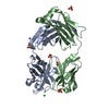 7ke1C 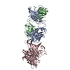 7ketC 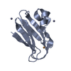 3cvdS C: citing same article ( S: Starting model for refinement |
|---|---|
| Similar structure data |


-Links
 PDBj
PDBj


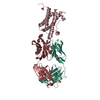
- Assembly
| Deposited unit | 
| ||||||||||||
|---|---|---|---|---|---|---|---|---|---|---|---|---|---|
| 1 |
| ||||||||||||
| Unit cell |
|
-Components
| #1: Antibody | Mass: 23978.697 Da / Num. of mol.: 1 Source method: isolated from a genetically manipulated source Source: (gene. exp.) Homo sapiens (human) / Production host:   Cricetulus griseus (Chinese hamster) Cricetulus griseus (Chinese hamster) | ||||
|---|---|---|---|---|---|
| #2: Antibody | Mass: 23565.068 Da / Num. of mol.: 1 Source method: isolated from a genetically manipulated source Source: (gene. exp.) Homo sapiens (human) / Production host: Cricetulus griseus (Chinese hamster) | ||||
| #3: Protein | Mass: 28074.248 Da / Num. of mol.: 1 Source method: isolated from a genetically manipulated source Source: (gene. exp.) Neisseria meningitidis (bacteria) / Gene: fhbp / Production host: Escherichia coli (E. coli) / References: UniProt: Q6VRZ6 | ||||
| #4: Chemical | Chloride  Mass: 35.453 Da / Num. of mol.: 2 / Source method: obtained synthetically / Formula: Cl Mass: 35.453 Da / Num. of mol.: 2 / Source method: obtained synthetically / Formula: Cl#5: Water | ChemComp-HOH / | Water Mass: 18.015 Da / Num. of mol.: 301 / Source method: isolated from a natural source / Formula: H2O Mass: 18.015 Da / Num. of mol.: 301 / Source method: isolated from a natural source / Formula: H2OHas ligand of interest | N | |
-Experimental details
-Experiment
| Experiment | Method: X-RAY DIFFRACTION / Number of used crystals: 1 |
|---|
- Sample preparation
| Crystal | Density Matthews: 2.13 Å3/Da / Density % sol: 42.38 % |
|---|---|
| Crystal grow | Temperature: 289 K / Method: vapor diffusion, hanging drop / pH: 7 / Details: 0.2 M LiNO3, 16% PEG 3350 |
-Data collection
| Diffraction | Mean temperature: 100 K / Serial crystal experiment: N |
|---|---|
| Diffraction source | Source: SYNCHROTRON / Site: ALS / Beamline: 8.3.1 / Wavelength: 1.11 Å |
| Detector | Type: DECTRIS PILATUS3 S 6M / Detector: PIXEL / Date: Sep 23, 2020 |
| Radiation | Monochromator: Si(111) / Protocol: SINGLE WAVELENGTH / Monochromatic (M) / Laue (L): M / Scattering type: x-ray |
| Radiation wavelength | Wavelength: 1.11 Å / Relative weight: 1 |
| Reflection | Resolution: 1.7→81.18 Å / Num. obs: 65915 / % possible obs: 93.1 % / Redundancy: 3.6 % / Biso Wilson estimate: 30.68 Å2 / CC1/2: 0.998 / CC star: 1 / Rmerge(I) obs: 0.052 / Rpim(I) all: 0.032 / Rrim(I) all: 0.061 / Net I/σ(I): 11.59 |
| Reflection shell | Resolution: 1.7→1.76 Å / Redundancy: 3.6 % / Rmerge(I) obs: 0.836 / Mean I/σ(I) obs: 1.3 / Num. unique obs: 6477 / CC1/2: 0.798 / CC star: 0.942 / Rpim(I) all: 0.513 / Rrim(I) all: 0.983 / % possible all: 93.1 |
- Processing
| Software |
| |||||||||||||||||||||||||||||||||||||||||||||||||||||||||||||||
|---|---|---|---|---|---|---|---|---|---|---|---|---|---|---|---|---|---|---|---|---|---|---|---|---|---|---|---|---|---|---|---|---|---|---|---|---|---|---|---|---|---|---|---|---|---|---|---|---|---|---|---|---|---|---|---|---|---|---|---|---|---|---|---|---|
| Refinement | Method to determine structure: MOLECULAR REPLACEMENT Starting model: 3CVD and Fab homology model Resolution: 1.7→81.18 Å / SU ML: 0.1784 / Cross valid method: FREE R-VALUE / σ(F): 1.96 / Phase error: 38.8457 Stereochemistry target values: GeoStd + Monomer Library + CDL v1.2
| |||||||||||||||||||||||||||||||||||||||||||||||||||||||||||||||
| Solvent computation | Shrinkage radii: 0.9 Å / VDW probe radii: 1.11 Å / Solvent model: FLAT BULK SOLVENT MODEL | |||||||||||||||||||||||||||||||||||||||||||||||||||||||||||||||
| Displacement parameters | Biso mean: 45.72 Å2 | |||||||||||||||||||||||||||||||||||||||||||||||||||||||||||||||
| Refinement step | Cycle: LAST / Resolution: 1.7→81.18 Å
| |||||||||||||||||||||||||||||||||||||||||||||||||||||||||||||||
| Refine LS restraints |
| |||||||||||||||||||||||||||||||||||||||||||||||||||||||||||||||
| LS refinement shell |
|
