 Movie
Movie Controller


+ Open data

- Basic information
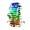
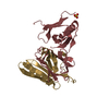
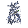
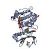
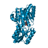
| Entry | Database: PDB / ID: 7lcm | ||||||
|---|---|---|---|---|---|---|---|
| Title | Receiver Domain of RssB bound to beryllofluoride | ||||||
 Components Components | Regulator of RpoS | ||||||
 Keywords Keywords |  SIGNALING PROTEIN / receiver domain / response regulator / ClpXP adaptor SIGNALING PROTEIN / receiver domain / response regulator / ClpXP adaptor | ||||||
| Function / homology |  Function and homology information Function and homology informationsigma factor antagonist activity / sigma factor antagonist complex / phosphorelay response regulator activity / positive regulation of proteolysis / protein-DNA complex / protein destabilization / transcription cis-regulatory region binding / negative regulation of DNA-templated transcription / regulation of DNA-templated transcription / cytosolSimilarity search - Function | ||||||
| Biological species |  Escherichia coli (E. coli) Escherichia coli (E. coli) | ||||||
| Method | X-RAY DIFFRACTION / SYNCHROTRON / MOLECULAR REPLACEMENT / Resolution: 1.91 Å | ||||||
 Authors Authors | Deaconescu, A.M. / Schwartz, J. / Son, J. | ||||||
| Funding support |  United States, 1items United States, 1items
| ||||||
 Citation Citation | Journal: Protein Sci. / Year: 2021 Title: Phospho-dependent signaling during the general stress response by the atypical response regulator and ClpXP adaptor RssB. Authors: Schwartz, J. / Son, J. / Brugger, C. / Deaconescu, A.M. | ||||||
| History |
|
- Structure visualization
| Structure viewer | Molecule: MolmilJmol/JSmol |
|---|
- Downloads & links
-Download
| PDBx/mmCIF format | 7lcm.cif.gz | 79.4 KB | Display | PDBx/mmCIF format |
|---|---|---|---|---|
| PDB format | pdb7lcm.ent.gz | 47.7 KB | Display | PDB format |
| PDBx/mmJSON format | 7lcm.json.gz | Tree view | PDBx/mmJSON format | |
| Others |  Other downloads Other downloads |
-Validation report
| Arichive directory | https://data.pdbj.org/pub/pdb/validation_reports/lc/7lcmftp://data.pdbj.org/pub/pdb/validation_reports/lc/7lcm | HTTPS FTP |
|---|
-Related structure data
| Related structure data |  7l9cSC S: Starting model for refinement C: citing same article ( |
|---|---|
| Similar structure data |


-Links
 PDBj
PDBj


- Assembly
| Deposited unit | 
| ||||||||||||
|---|---|---|---|---|---|---|---|---|---|---|---|---|---|
| 1 | 
| ||||||||||||
| Unit cell |
| ||||||||||||
| Components on special symmetry positions |
|
-Components
| #1: Protein | Mass: 14566.069 Da / Num. of mol.: 1 Source method: isolated from a genetically manipulated source Source: (gene. exp.) Escherichia coli (E. coli) / Strain: MG1655 / Gene: rssB / Production host: Escherichia coli (E. coli) / References: UniProt: P0AEV1 |
|---|---|
| #2: Chemical | ChemComp-MG /   Mass: 24.305 Da / Num. of mol.: 1 / Source method: obtained synthetically / Formula: Mg / Feature type: SUBJECT OF INVESTIGATION Mass: 24.305 Da / Num. of mol.: 1 / Source method: obtained synthetically / Formula: Mg / Feature type: SUBJECT OF INVESTIGATION |
| #3: Chemical | ChemComp-BEF /   Mass: 66.007 Da / Num. of mol.: 1 / Source method: obtained synthetically / Formula: BeF3 / Feature type: SUBJECT OF INVESTIGATION Mass: 66.007 Da / Num. of mol.: 1 / Source method: obtained synthetically / Formula: BeF3 / Feature type: SUBJECT OF INVESTIGATION |
| #4: Water | ChemComp-HOH / Water Mass: 18.015 Da / Num. of mol.: 44 / Source method: isolated from a natural source / Formula: H2O Mass: 18.015 Da / Num. of mol.: 44 / Source method: isolated from a natural source / Formula: H2O |
| Has ligand of interest | Y |
-Experimental details
-Experiment
| Experiment | Method: X-RAY DIFFRACTION / Number of used crystals: 1 |
|---|
- Sample preparation
| Crystal | Density Matthews: 2.1 Å3/Da / Density % sol: 41.44 % |
|---|---|
| Crystal grow | Temperature: 298 K / Method: vapor diffusion, sitting drop / pH: 8.8 Details: 0.2M lithium sulfate, 22% PEG 3,350, and 0.1M Tris-HCl pH 8.8 |
-Data collection
| Diffraction | Mean temperature: 100 K / Serial crystal experiment: N |
|---|---|
| Diffraction source | Source: SYNCHROTRON / Site: APS / Beamline: 24-ID-C / Wavelength: 0.97918 Å |
| Detector | Type: DECTRIS EIGER2 X 16M / Detector: PIXEL / Date: Apr 12, 2020 |
| Radiation | Protocol: SINGLE WAVELENGTH / Monochromatic (M) / Laue (L): M / Scattering type: x-ray |
| Radiation wavelength | Wavelength: 0.97918 Å / Relative weight: 1 |
| Reflection | Resolution: 1.9→40.12 Å / Num. obs: 9736 / % possible obs: 99.5 % / Redundancy: 9.3 % / Biso Wilson estimate: 35.97 Å2 / CC1/2: 0.999 / Rpim(I) all: 0.028 / Net I/σ(I): 21 |
| Reflection shell | Resolution: 1.91→1.95 Å / Mean I/σ(I) obs: 1.7 / Num. unique obs: 908 / CC1/2: 0.69 / Rpim(I) all: 0.366 |
- Processing
| Software |
| ||||||||||||||||||||||||||||||||||||||||||||||||||||||||
|---|---|---|---|---|---|---|---|---|---|---|---|---|---|---|---|---|---|---|---|---|---|---|---|---|---|---|---|---|---|---|---|---|---|---|---|---|---|---|---|---|---|---|---|---|---|---|---|---|---|---|---|---|---|---|---|---|---|
| Refinement | Method to determine structure: MOLECULAR REPLACEMENT Starting model: 7L9C Resolution: 1.91→40.12 Å / SU ML: 0.1592 / Cross valid method: FREE R-VALUE / σ(F): 1.35 / Phase error: 29.1339 Stereochemistry target values: GeoStd + Monomer Library + CDL v1.2
| ||||||||||||||||||||||||||||||||||||||||||||||||||||||||
| Solvent computation | Shrinkage radii: 0.9 Å / VDW probe radii: 1.11 Å / Solvent model: FLAT BULK SOLVENT MODEL | ||||||||||||||||||||||||||||||||||||||||||||||||||||||||
| Displacement parameters | Biso mean: 44.01 Å2 | ||||||||||||||||||||||||||||||||||||||||||||||||||||||||
| Refinement step | Cycle: LAST / Resolution: 1.91→40.12 Å
| ||||||||||||||||||||||||||||||||||||||||||||||||||||||||
| Refine LS restraints |
| ||||||||||||||||||||||||||||||||||||||||||||||||||||||||
| LS refinement shell |
| ||||||||||||||||||||||||||||||||||||||||||||||||||||||||
| Refinement TLS params. | Method: refined / Origin x: 12.0058862267 Å / Origin y: -10.5907234563 Å / Origin z: 9.23832018953 Å
| ||||||||||||||||||||||||||||||||||||||||||||||||||||||||
| Refinement TLS group | Selection details: all |
