 Movie
Movie Controller


+ Open data

- Basic information
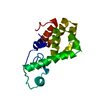
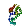
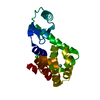
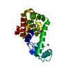
| Entry | Database: PDB / ID: 7l3a | ||||||
|---|---|---|---|---|---|---|---|
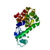
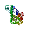
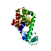
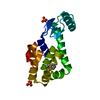
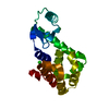
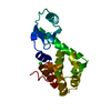
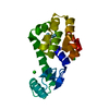
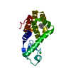
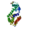
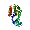
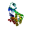
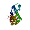
| Title | T4 Lysozyme L99A - toluene - cryo | ||||||
 Components Components | Endolysin Lysin Lysin | ||||||
 Keywords Keywords | HYDROLASE / cryo / toluene / L99A | ||||||
| Function / homology |  Function and homology information Function and homology informationviral release from host cell by cytolysis / peptidoglycan catabolic process / cell wall macromolecule catabolic process / lysozyme / lysozyme activity / host cell cytoplasm / defense response to bacteriumSimilarity search - Function | ||||||
| Biological species |  Enterobacteria phage T4 (virus) Enterobacteria phage T4 (virus) | ||||||
| Method | X-RAY DIFFRACTION / SYNCHROTRON / MOLECULAR REPLACEMENT / molecular replacement / Resolution: 1.11 Å | ||||||
 Authors Authors | Fischer, M. / Bradford, S.Y.C. | ||||||
 Citation Citation | Journal: Chem Sci / Year: 2021 Title: Temperature artifacts in protein structures bias ligand-binding predictions. Authors: Bradford, S.Y.C. / El Khoury, L. / Ge, Y. / Osato, M. / Mobley, D.L. / Fischer, M. | ||||||
| History |
|
- Structure visualization
| Structure viewer | Molecule: MolmilJmol/JSmol |
|---|
- Downloads & links
-Download
| PDBx/mmCIF format | 7l3a.cif.gz | 86.6 KB | Display | PDBx/mmCIF format |
|---|---|---|---|---|
| PDB format | pdb7l3a.ent.gz | 63.8 KB | Display | PDB format |
| PDBx/mmJSON format | 7l3a.json.gz | Tree view | PDBx/mmJSON format | |
| Others |  Other downloads Other downloads |
-Validation report
| Arichive directory | https://data.pdbj.org/pub/pdb/validation_reports/l3/7l3aftp://data.pdbj.org/pub/pdb/validation_reports/l3/7l3a | HTTPS FTP |
|---|
-Related structure data
| Related structure data |  7l37C  7l38C  7l39C 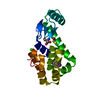 7l3bC 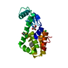 7l3cC  7l3dC 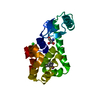 7l3eC  7l3fC 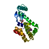 7l3gC 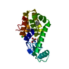 7l3hC  7l3iC  7l3jC 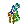 7l3kC  4w51S S: Starting model for refinement C: citing same article ( |
|---|---|
| Similar structure data |


-Links
 PDBj
PDBj


- Assembly
| Deposited unit | 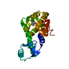
| ||||||||
|---|---|---|---|---|---|---|---|---|---|
| 1 |
| ||||||||
| Unit cell |
| ||||||||
| Components on special symmetry positions |
|
-Components
| #1: Protein | Lysin / Lysis protein / Lysozyme / Muramidase Mass: 18620.387 Da / Num. of mol.: 1 / Mutation: R12G/I137R/L99A Source method: isolated from a genetically manipulated source Source: (gene. exp.) Enterobacteria phage T4 (virus) / Production host:  Escherichia coli (E. coli) / References: UniProt: P00720, lysozyme Escherichia coli (E. coli) / References: UniProt: P00720, lysozyme |
|---|---|
| #2: Chemical | ChemComp-MBN / Toluene  Mass: 92.138 Da / Num. of mol.: 1 / Source method: obtained synthetically / Formula: C7H8 / Feature type: SUBJECT OF INVESTIGATION Mass: 92.138 Da / Num. of mol.: 1 / Source method: obtained synthetically / Formula: C7H8 / Feature type: SUBJECT OF INVESTIGATION |
| #3: Water | ChemComp-HOH / Water Mass: 18.015 Da / Num. of mol.: 165 / Source method: isolated from a natural source / Formula: H2O Mass: 18.015 Da / Num. of mol.: 165 / Source method: isolated from a natural source / Formula: H2O |
| Has ligand of interest | Y |
-Experimental details
-Experiment
| Experiment | Method: X-RAY DIFFRACTION / Number of used crystals: 1 |
|---|
- Sample preparation
| Crystal | Density Matthews: 2.68 Å3/Da / Density % sol: 54.07 % |
|---|---|
| Crystal grow | Temperature: 291 K / Method: vapor diffusion, hanging drop Details: Crystals were grown from a 10 mg/mL frozen protein solution by the hanging drop method at 291-293K, with a 1:1 drop ratio of protein to solution and over a well solution of 0.1 M Tris- ...Details: Crystals were grown from a 10 mg/mL frozen protein solution by the hanging drop method at 291-293K, with a 1:1 drop ratio of protein to solution and over a well solution of 0.1 M Tris-hydrochloride (pH 8), 20%-26% (w/v) PEG 4000, 70-170 mM lithium citrate, 8%-18% 2-propanol, 50 mM 2-mercaptoethanol, and 50 mM 2-hydroxyethyl disulfide Temp details: 291-293 |
-Data collection
| Diffraction | Mean temperature: 100 K / Serial crystal experiment: N | ||||||||||||||||||
|---|---|---|---|---|---|---|---|---|---|---|---|---|---|---|---|---|---|---|---|
| Diffraction source | Source: SYNCHROTRON / Site: APS  / Beamline: 22-ID / Wavelength: 1 Å / Beamline: 22-ID / Wavelength: 1 Å | ||||||||||||||||||
| Detector | Type: DECTRIS EIGER X 16M / Detector: PIXEL / Date: Feb 14, 2018 | ||||||||||||||||||
| Radiation | Protocol: SINGLE WAVELENGTH / Monochromatic (M) / Laue (L): M / Scattering type: x-ray | ||||||||||||||||||
| Radiation wavelength | Wavelength: 1 Å / Relative weight: 1 | ||||||||||||||||||
| Reflection | Resolution: 1.11→45.67 Å / Num. obs: 74260 / % possible obs: 96 % / Redundancy: 8.8 % / Rmerge(I) obs: 0.049 / Net I/σ(I): 16.2 | ||||||||||||||||||
| Reflection shell |
|
-Phasing
| Phasing | Method: molecular replacement | |||||||||
|---|---|---|---|---|---|---|---|---|---|---|
| Phasing MR |
|
- Processing
| Software |
| ||||||||||||||||||||||||||||||||||||||||||||||||||||||||||||||||||||||||||||||||||||||||||
|---|---|---|---|---|---|---|---|---|---|---|---|---|---|---|---|---|---|---|---|---|---|---|---|---|---|---|---|---|---|---|---|---|---|---|---|---|---|---|---|---|---|---|---|---|---|---|---|---|---|---|---|---|---|---|---|---|---|---|---|---|---|---|---|---|---|---|---|---|---|---|---|---|---|---|---|---|---|---|---|---|---|---|---|---|---|---|---|---|---|---|---|
| Refinement | Method to determine structure: MOLECULAR REPLACEMENT Starting model: 4w51 Resolution: 1.11→45.67 Å / SU ML: 0.1 / Cross valid method: THROUGHOUT / σ(F): 1.35 / Phase error: 23.27 / Stereochemistry target values: ML
| ||||||||||||||||||||||||||||||||||||||||||||||||||||||||||||||||||||||||||||||||||||||||||
| Solvent computation | Shrinkage radii: 0.9 Å / VDW probe radii: 1.11 Å / Solvent model: FLAT BULK SOLVENT MODEL | ||||||||||||||||||||||||||||||||||||||||||||||||||||||||||||||||||||||||||||||||||||||||||
| Displacement parameters | Biso max: 50.46 Å2 / Biso mean: 15.9005 Å2 / Biso min: 8.97 Å2 | ||||||||||||||||||||||||||||||||||||||||||||||||||||||||||||||||||||||||||||||||||||||||||
| Refinement step | Cycle: final / Resolution: 1.11→45.67 Å
| ||||||||||||||||||||||||||||||||||||||||||||||||||||||||||||||||||||||||||||||||||||||||||
| Refine LS restraints |
| ||||||||||||||||||||||||||||||||||||||||||||||||||||||||||||||||||||||||||||||||||||||||||
| LS refinement shell | Refine-ID: X-RAY DIFFRACTION / Rfactor Rfree error: 0
|
