 Movie
Movie Controller

[English] 日本語
 Yorodumi
- PDB-7l1c: Crystal structure of HLA-A*03:01 in complex with a mutant PIK3CA ... -
+ Open data

- Basic information
| Entry | Database: PDB / ID: 7l1c | ||||||
|---|---|---|---|---|---|---|---|
| Title | Crystal structure of HLA-A*03:01 in complex with a mutant PIK3CA peptide | ||||||
 Components Components |
| ||||||
 Keywords Keywords |  IMMUNE SYSTEM / peptide major histocompatibility complex IMMUNE SYSTEM / peptide major histocompatibility complex | ||||||
| Function / homology |  Function and homology information Function and homology informationresponse to muscle inactivity / negative regulation of actin filament depolymerization / response to L-leucine / regulation of actin filament organization / response to butyrate / autosome genomic imprinting / IRS-mediated signalling / cellular response to hydrostatic pressure / PI3K events in ERBB4 signaling / Activated NTRK2 signals through PI3K ...response to muscle inactivity / negative regulation of actin filament depolymerization / response to L-leucine / regulation of actin filament organization / response to butyrate / autosome genomic imprinting / IRS-mediated signalling / cellular response to hydrostatic pressure / PI3K events in ERBB4 signaling / Activated NTRK2 signals through PI3K / positive regulation of protein localization to membrane / Activated NTRK3 signals through PI3K / negative regulation of fibroblast apoptotic process / cardiac muscle cell contraction / phosphatidylinositol 3-kinase complex, class IB / vasculature development / Signaling by cytosolic FGFR1 fusion mutants / regulation of cellular respiration / phosphatidylinositol 3-kinase complex / anoikis / Nephrin family interactions / 1-phosphatidylinositol-4-phosphate 3-kinase activity / Costimulation by the CD28 family / vascular endothelial growth factor signaling pathway / 1-phosphatidylinositol-4,5-bisphosphate 3-kinase activity / MET activates PI3K/AKT signaling / PI3K/AKT activation / phosphatidylinositol-4,5-bisphosphate 3-kinase / phosphatidylinositol 3-kinase complex, class IA / phosphatidylinositol 3-kinase / relaxation of cardiac muscle / phosphatidylinositol-3-phosphate biosynthetic process / 1-phosphatidylinositol-3-kinase activity / negative regulation of macroautophagy / Signaling by ALK / PI-3K cascade:FGFR3 / Erythropoietin activates Phosphoinositide-3-kinase (PI3K) / protein kinase activator activity / response to dexamethasone / PI-3K cascade:FGFR2 / PI-3K cascade:FGFR4 / PI-3K cascade:FGFR1 / CD28 dependent PI3K/Akt signaling / Synthesis of PIPs at the plasma membrane / phosphatidylinositol phosphate biosynthetic process / T cell mediated cytotoxicity directed against tumor cell target / antigen processing and presentation of endogenous peptide antigen via MHC class I via ER pathway, TAP-dependent / positive regulation of memory T cell activation / TAP complex binding / PI3K events in ERBB2 signaling / Signaling by ALK fusions and activated point mutants / antigen processing and presentation of exogenous peptide antigen via MHC class I / Golgi medial cisterna / negative regulation of anoikis / RET signaling / regulation of multicellular organism growth / insulin receptor substrate binding / positive regulation of CD8-positive, alpha-beta T cell activation / Interleukin-3, Interleukin-5 and GM-CSF signaling / CD8-positive, alpha-beta T cell activation / PI3K Cascade / intercalated disc / positive regulation of TOR signaling / positive regulation of CD8-positive, alpha-beta T cell proliferation / CD8 receptor binding / endothelial cell migration / RAC2 GTPase cycle / GAB1 signalosome / Role of phospholipids in phagocytosis / Role of LAT2/NTAL/LAB on calcium mobilization / adipose tissue development / Interleukin receptor SHC signaling / endoplasmic reticulum exit site / beta-2-microglobulin binding / positive regulation of lamellipodium assembly / phagocytosis / Signaling by PDGFRA transmembrane, juxtamembrane and kinase domain mutants / Signaling by PDGFRA extracellular domain mutants / energy homeostasis / Signaling by FGFR4 in disease / TAP binding / Signaling by FLT3 ITD and TKD mutants / protection from natural killer cell mediated cytotoxicity / cardiac muscle contraction / Signaling by FGFR3 in disease / GPVI-mediated activation cascade / Tie2 Signaling / antigen processing and presentation of endogenous peptide antigen via MHC class I via ER pathway, TAP-independent / antigen processing and presentation of endogenous peptide antigen via MHC class Ib / Signaling by FGFR2 in disease / RAC1 GTPase cycle / T cell costimulation / response to muscle stretch / Signaling by FLT3 fusion proteins / detection of bacterium / FLT3 Signaling / Signaling by FGFR1 in disease / T cell receptor binding / Downstream signal transduction / insulin-like growth factor receptor signaling pathwaySimilarity search - Function | ||||||
| Biological species |  Homo sapiens (human) Homo sapiens (human) | ||||||
| Method | X-RAY DIFFRACTION / SYNCHROTRON / MOLECULAR REPLACEMENT / Resolution: 1.96 Å | ||||||
 Authors Authors | Ma, J. / Baker, B.M. | ||||||
| Funding support |  United States, 1items United States, 1items
| ||||||
 Citation Citation | Journal: Nat Med / Year: 2022 Title: Immunogenicity and therapeutic targeting of a public neoantigen derived from mutated PIK3CA. Authors: Chandran, S.S. / Ma, J. / Klatt, M.G. / Dundar, F. / Bandlamudi, C. / Razavi, P. / Wen, H.Y. / Weigelt, B. / Zumbo, P. / Fu, S.N. / Banks, L.B. / Yi, F. / Vercher, E. / Etxeberria, I. / ...Authors: Chandran, S.S. / Ma, J. / Klatt, M.G. / Dundar, F. / Bandlamudi, C. / Razavi, P. / Wen, H.Y. / Weigelt, B. / Zumbo, P. / Fu, S.N. / Banks, L.B. / Yi, F. / Vercher, E. / Etxeberria, I. / Bestman, W.D. / Da Cruz Paula, A. / Aricescu, I.S. / Drilon, A. / Betel, D. / Scheinberg, D.A. / Baker, B.M. / Klebanoff, C.A. | ||||||
| History |
|
- Structure visualization
| Structure viewer | Molecule: MolmilJmol/JSmol |
|---|
- Downloads & links
-Download
| PDBx/mmCIF format | 7l1c.cif.gz | 213.4 KB | Display | PDBx/mmCIF format |
|---|---|---|---|---|
| PDB format | pdb7l1c.ent.gz | 141.9 KB | Display | PDB format |
| PDBx/mmJSON format | 7l1c.json.gz | Tree view | PDBx/mmJSON format | |
| Others |  Other downloads Other downloads |
-Validation report
| Arichive directory | https://data.pdbj.org/pub/pdb/validation_reports/l1/7l1cftp://data.pdbj.org/pub/pdb/validation_reports/l1/7l1c | HTTPS FTP |
|---|
-Related structure data
| Related structure data | 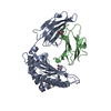 7l1bC 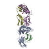 7l1dC 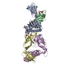 7rrgC 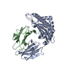 2xpgS S: Starting model for refinement C: citing same article ( |
|---|---|
| Similar structure data |
-Links
 PDBj
PDBj


- Assembly
| Deposited unit | 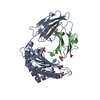
| ||||||||||||
|---|---|---|---|---|---|---|---|---|---|---|---|---|---|
| 1 |
| ||||||||||||
| Unit cell |
|
-Components
-Protein , 2 types, 2 molecules AB
| #1: Protein | Mass: 31628.838 Da / Num. of mol.: 1 Source method: isolated from a genetically manipulated source Source: (gene. exp.) Homo sapiens (human) / Gene: HLA-A, HLAA / Production host:  Escherichia coli (E. coli) / References: UniProt: P04439 Escherichia coli (E. coli) / References: UniProt: P04439 |
|---|---|
| #2: Protein | Beta-2 microglobulin Mass: 11879.356 Da / Num. of mol.: 1 Source method: isolated from a genetically manipulated source Source: (gene. exp.) Homo sapiens (human) / Gene: B2M, CDABP0092, HDCMA22P / Production host: Escherichia coli (E. coli) / References: UniProt: P61769 |
-Protein/peptide , 1 types, 1 molecules C
| #3: Protein/peptide | Mass: 972.098 Da / Num. of mol.: 1 / Source method: obtained synthetically / Source: (synth.) Homo sapiens (human) / References: UniProt: P42336 |
|---|
-Non-polymers , 3 types, 323 molecules 


| #4: Chemical | ChemComp-GOL / Glycerol Mass: 92.094 Da / Num. of mol.: 5 / Source method: obtained synthetically / Formula: C3H8O3 / Feature type: SUBJECT OF INVESTIGATION Mass: 92.094 Da / Num. of mol.: 5 / Source method: obtained synthetically / Formula: C3H8O3 / Feature type: SUBJECT OF INVESTIGATION#5: Chemical | Formic acid Mass: 46.025 Da / Num. of mol.: 3 / Source method: obtained synthetically / Formula: CH2O2 / Feature type: SUBJECT OF INVESTIGATION Mass: 46.025 Da / Num. of mol.: 3 / Source method: obtained synthetically / Formula: CH2O2 / Feature type: SUBJECT OF INVESTIGATION#6: Water | ChemComp-HOH / | WaterMass: 18.015 Da / Num. of mol.: 315 / Source method: isolated from a natural source / Formula: H2O |
|---|
-Details
| Has ligand of interest | Y |
|---|
-Experimental details
-Experiment
| Experiment | Method: X-RAY DIFFRACTION / Number of used crystals: 1 |
|---|
- Sample preparation
| Crystal | Density Matthews: 3.38 Å3/Da / Density % sol: 63.57 % |
|---|---|
| Crystal grow | Temperature: 277.15 K / Method: vapor diffusion, hanging drop / pH: 6.6 Details: 20% w/v Polyethylene glycol 3,350, 200 mM Ammonium formate, pH 6.6 |
-Data collection
| Diffraction | Mean temperature: 100 K / Serial crystal experiment: N |
|---|---|
| Diffraction source | Source: SYNCHROTRON / Site: APS / Beamline: 24-ID-E / Wavelength: 1 Å |
| Detector | Type: DECTRIS EIGER X 16M / Detector: PIXEL / Date: Apr 2, 2019 |
| Radiation | Protocol: SINGLE WAVELENGTH / Monochromatic (M) / Laue (L): M / Scattering type: x-ray |
| Radiation wavelength | Wavelength: 1 Å / Relative weight: 1 |
| Reflection | Resolution: 1.96→50 Å / Num. obs: 44346 / % possible obs: 99.86 % / Redundancy: 17.6 % / Biso Wilson estimate: 25.5 Å2 / Rmerge(I) obs: 0.123 / Rpim(I) all: 0.029 / Rrim(I) all: 0.127 / Net I/σ(I): 29.3 |
| Reflection shell | Resolution: 1.96→1.99 Å / Redundancy: 10.9 % / Rmerge(I) obs: 0.918 / Num. unique obs: 2187 / CC1/2: 0.8 / Rpim(I) all: 0.267 / Rrim(I) all: 0.96 / % possible all: 100 |
- Processing
| Software |
| |||||||||||||||||||||||||||||||||||||||||||||||||||||||||||||||||||||||||||||||||||||||||||||||||||||||||||||||||||||||||||||
|---|---|---|---|---|---|---|---|---|---|---|---|---|---|---|---|---|---|---|---|---|---|---|---|---|---|---|---|---|---|---|---|---|---|---|---|---|---|---|---|---|---|---|---|---|---|---|---|---|---|---|---|---|---|---|---|---|---|---|---|---|---|---|---|---|---|---|---|---|---|---|---|---|---|---|---|---|---|---|---|---|---|---|---|---|---|---|---|---|---|---|---|---|---|---|---|---|---|---|---|---|---|---|---|---|---|---|---|---|---|---|---|---|---|---|---|---|---|---|---|---|---|---|---|---|---|---|
| Refinement | Method to determine structure: MOLECULAR REPLACEMENT Starting model: 2XPG Resolution: 1.96→44.95 Å / SU ML: 0.2236 / Cross valid method: FREE R-VALUE / σ(F): 0 / Phase error: 19.7119 Stereochemistry target values: GeoStd + Monomer Library + CDL v1.2
| |||||||||||||||||||||||||||||||||||||||||||||||||||||||||||||||||||||||||||||||||||||||||||||||||||||||||||||||||||||||||||||
| Solvent computation | Shrinkage radii: 0.9 Å / VDW probe radii: 1.11 Å / Solvent model: FLAT BULK SOLVENT MODEL | |||||||||||||||||||||||||||||||||||||||||||||||||||||||||||||||||||||||||||||||||||||||||||||||||||||||||||||||||||||||||||||
| Displacement parameters | Biso mean: 37.12 Å2 | |||||||||||||||||||||||||||||||||||||||||||||||||||||||||||||||||||||||||||||||||||||||||||||||||||||||||||||||||||||||||||||
| Refinement step | Cycle: LAST / Resolution: 1.96→44.95 Å
| |||||||||||||||||||||||||||||||||||||||||||||||||||||||||||||||||||||||||||||||||||||||||||||||||||||||||||||||||||||||||||||
| Refine LS restraints |
| |||||||||||||||||||||||||||||||||||||||||||||||||||||||||||||||||||||||||||||||||||||||||||||||||||||||||||||||||||||||||||||
| LS refinement shell |
| |||||||||||||||||||||||||||||||||||||||||||||||||||||||||||||||||||||||||||||||||||||||||||||||||||||||||||||||||||||||||||||
| Refinement TLS params. | Method: refined / Refine-ID: X-RAY DIFFRACTION
| |||||||||||||||||||||||||||||||||||||||||||||||||||||||||||||||||||||||||||||||||||||||||||||||||||||||||||||||||||||||||||||
| Refinement TLS group |
|
