 Movie
Movie Controller


+ Open data

- Basic information
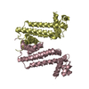
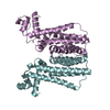
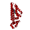
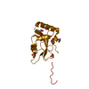
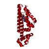
| Entry | Database: PDB / ID: 7kji | |||||||||
|---|---|---|---|---|---|---|---|---|---|---|
| Title | Plasmodium falciparum protein Pf12p bound to nanobody D9 | |||||||||
 Components Components |
| |||||||||
 Keywords Keywords | UNKNOWN FUNCTION /  Plasmodium falciparum / 6-cysteine protein / s48/45 domain / nanobody Plasmodium falciparum / 6-cysteine protein / s48/45 domain / nanobody | |||||||||
| Function / homology | 6-Cysteine (6-Cys) domain / 6-Cysteine (6-Cys) domain superfamily / Sexual stage antigen s48/45 domain / 6-Cysteine (6-Cys) domain profile. / Sexual stage antigen s48/45 domain / cell surface / plasma membrane / Surface protein P12p Function and homology information Function and homology information | |||||||||
| Biological species |  Vicugna pacos (alpaca) Vicugna pacos (alpaca) Plasmodium falciparum (malaria parasite P. falciparum) Plasmodium falciparum (malaria parasite P. falciparum) | |||||||||
| Method | X-RAY DIFFRACTION / SYNCHROTRON / MOLECULAR REPLACEMENT / Resolution: 3.25 Å | |||||||||
 Authors Authors | Dietrich, M.H. / Tham, W.H. | |||||||||
| Funding support |  Australia, Australia,  United Kingdom, 2items United Kingdom, 2items
| |||||||||
 Citation Citation | Journal: Biochem.J. / Year: 2021 Title: Nanobody generation and structural characterization of Plasmodium falciparum 6-cysteine protein Pf12p. Authors: Dietrich, M.H. / Chan, L.J. / Adair, A. / Keremane, S. / Pymm, P. / Lo, A.W. / Cao, Y.C. / Tham, W.H. | |||||||||
| History |
|
- Structure visualization
| Structure viewer | Molecule: MolmilJmol/JSmol |
|---|
- Downloads & links
-Download
| PDBx/mmCIF format | 7kji.cif.gz | 87 KB | Display | PDBx/mmCIF format |
|---|---|---|---|---|
| PDB format | pdb7kji.ent.gz | 61.2 KB | Display | PDB format |
| PDBx/mmJSON format | 7kji.json.gz | Tree view | PDBx/mmJSON format | |
| Others |  Other downloads Other downloads |
-Validation report
| Arichive directory | https://data.pdbj.org/pub/pdb/validation_reports/kj/7kjiftp://data.pdbj.org/pub/pdb/validation_reports/kj/7kji | HTTPS FTP |
|---|
-Related structure data
| Related structure data |  7kj7SC 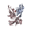 7kjhC S: Starting model for refinement C: citing same article ( |
|---|---|
| Similar structure data |


-Links
 PDBj
PDBj


- Assembly
| Deposited unit | 
| ||||||||
|---|---|---|---|---|---|---|---|---|---|
| 1 |
| ||||||||
| Unit cell |
|
-Components
| #1: Antibody | Mass: 13642.261 Da / Num. of mol.: 1 Source method: isolated from a genetically manipulated source Source: (gene. exp.) Vicugna pacos (alpaca) / Production host:  Escherichia coli (E. coli) Escherichia coli (E. coli) |
|---|---|
| #2: Protein | Mass: 37247.488 Da / Num. of mol.: 1 Source method: isolated from a genetically manipulated source Source: (gene. exp.) Plasmodium falciparum (malaria parasite P. falciparum)Strain: isolate 3D7 / Gene: PFS12P, PF12P, PFF0620c / Production host:   Spodoptera frugiperda (fall armyworm) / References: UniProt: C6KSX1 Spodoptera frugiperda (fall armyworm) / References: UniProt: C6KSX1 |
-Experimental details
-Experiment
| Experiment | Method: X-RAY DIFFRACTION / Number of used crystals: 1 |
|---|
- Sample preparation
| Crystal | Density Matthews: 2.27 Å3/Da / Density % sol: 45.74 % |
|---|---|
| Crystal grow | Temperature: 281 K / Method: vapor diffusion, hanging drop / pH: 6.5 Details: 0.1 M bis-tris chloride pH 6.5, 0.2 M magnesium chloride, 25% PEG3350 |
-Data collection
| Diffraction | Mean temperature: 100 K / Serial crystal experiment: N | ||||||||||||||||||||||||||||||||||||||||||||||||||||||||||||||||||||||||||||||||||||||||||||||||||||
|---|---|---|---|---|---|---|---|---|---|---|---|---|---|---|---|---|---|---|---|---|---|---|---|---|---|---|---|---|---|---|---|---|---|---|---|---|---|---|---|---|---|---|---|---|---|---|---|---|---|---|---|---|---|---|---|---|---|---|---|---|---|---|---|---|---|---|---|---|---|---|---|---|---|---|---|---|---|---|---|---|---|---|---|---|---|---|---|---|---|---|---|---|---|---|---|---|---|---|---|---|---|
| Diffraction source | Source: SYNCHROTRON / Site: Australian Synchrotron / Beamline: MX2 / Wavelength: 0.953649 Å | ||||||||||||||||||||||||||||||||||||||||||||||||||||||||||||||||||||||||||||||||||||||||||||||||||||
| Detector | Type: DECTRIS EIGER X 16M / Detector: PIXEL / Date: Sep 22, 2019 | ||||||||||||||||||||||||||||||||||||||||||||||||||||||||||||||||||||||||||||||||||||||||||||||||||||
| Radiation | Protocol: SINGLE WAVELENGTH / Monochromatic (M) / Laue (L): M / Scattering type: x-ray | ||||||||||||||||||||||||||||||||||||||||||||||||||||||||||||||||||||||||||||||||||||||||||||||||||||
| Radiation wavelength | Wavelength: 0.953649 Å / Relative weight: 1 | ||||||||||||||||||||||||||||||||||||||||||||||||||||||||||||||||||||||||||||||||||||||||||||||||||||
| Reflection | Resolution: 3.25→42.753 Å / Num. obs: 7710 / % possible obs: 99.8 % / Redundancy: 13.102 % / Biso Wilson estimate: 99.233 Å2 / CC1/2: 0.999 / Rmerge(I) obs: 0.153 / Rrim(I) all: 0.16 / Χ2: 0.764 / Net I/σ(I): 13.51 / Num. measured all: 101019 | ||||||||||||||||||||||||||||||||||||||||||||||||||||||||||||||||||||||||||||||||||||||||||||||||||||
| Reflection shell | Diffraction-ID: 1
|
- Processing
| Software |
| ||||||||||||||||||||||||||||||||||||||||||||||||||||||||||||
|---|---|---|---|---|---|---|---|---|---|---|---|---|---|---|---|---|---|---|---|---|---|---|---|---|---|---|---|---|---|---|---|---|---|---|---|---|---|---|---|---|---|---|---|---|---|---|---|---|---|---|---|---|---|---|---|---|---|---|---|---|---|
| Refinement | Method to determine structure: MOLECULAR REPLACEMENT Starting model: 7KJ7 Resolution: 3.25→42.753 Å / SU ML: 0.48 / Cross valid method: THROUGHOUT / σ(F): 1.36 / Phase error: 31.61 / Stereochemistry target values: ML
| ||||||||||||||||||||||||||||||||||||||||||||||||||||||||||||
| Solvent computation | Shrinkage radii: 0.9 Å / VDW probe radii: 1.11 Å / Solvent model: FLAT BULK SOLVENT MODEL | ||||||||||||||||||||||||||||||||||||||||||||||||||||||||||||
| Displacement parameters | Biso max: 162.91 Å2 / Biso mean: 110.5409 Å2 / Biso min: 69.41 Å2 | ||||||||||||||||||||||||||||||||||||||||||||||||||||||||||||
| Refinement step | Cycle: final / Resolution: 3.25→42.753 Å
| ||||||||||||||||||||||||||||||||||||||||||||||||||||||||||||
| Refine LS restraints |
| ||||||||||||||||||||||||||||||||||||||||||||||||||||||||||||
| LS refinement shell | Refine-ID: X-RAY DIFFRACTION / Rfactor Rfree error: 0 / % reflection obs: 100 %
|
