Movie
Movie Controller


+ Open data

- Basic information
| Entry | Database: PDB / ID: 7kc4 | |||||||||
|---|---|---|---|---|---|---|---|---|---|---|
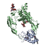
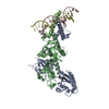
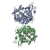
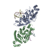
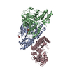
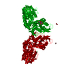
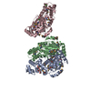
| Title | Human WLS in complex with WNT8A | |||||||||
 Components Components |
| |||||||||
 Keywords Keywords |  MEMBRANE PROTEIN / G-protein coupled receptor / palmitoleation / secretion / embryonic development MEMBRANE PROTEIN / G-protein coupled receptor / palmitoleation / secretion / embryonic development | |||||||||
| Function / homology |  Function and homology information: / : / Wnt protein secretion / neural crest cell fate commitment / positive regulation of Wnt protein secretion / WNT ligand biogenesis and trafficking / secondary palate development / cementum mineralization / beta-catenin destruction complex disassembly / hindbrain development ...: / : / Wnt protein secretion / neural crest cell fate commitment / positive regulation of Wnt protein secretion / WNT ligand biogenesis and trafficking / secondary palate development / cementum mineralization / beta-catenin destruction complex disassembly / hindbrain development / Wnt-protein binding / exocrine pancreas development / frizzled binding / Class B/2 (Secretin family receptors) / Disassembly of the destruction complex and recruitment of AXIN to the membrane / anterior/posterior axis specification / midbrain development / mesoderm formation / positive regulation of Wnt signaling pathway / canonical Wnt signaling pathway / endomembrane system / cell fate commitment / response to retinoic acid / TCF dependent signaling in response to WNT / cytokine activity / intracellular protein transport / trans-Golgi network / neuron differentiation / Wnt signaling pathway / positive regulation of canonical Wnt signaling pathway / endocytic vesicle membrane / cytoplasmic vesicle / early endosome membrane / collagen-containing extracellular matrix / positive regulation of canonical NF-kappaB signal transduction / receptor ligand activity / early endosome / Golgi membrane / endoplasmic reticulum membrane / Golgi apparatus / endoplasmic reticulum / positive regulation of transcription by RNA polymerase II / extracellular space / extracellular exosome / extracellular region / identical protein binding / plasma membrane / cytosol Function and homology information: / : / Wnt protein secretion / neural crest cell fate commitment / positive regulation of Wnt protein secretion / WNT ligand biogenesis and trafficking / secondary palate development / cementum mineralization / beta-catenin destruction complex disassembly / hindbrain development ...: / : / Wnt protein secretion / neural crest cell fate commitment / positive regulation of Wnt protein secretion / WNT ligand biogenesis and trafficking / secondary palate development / cementum mineralization / beta-catenin destruction complex disassembly / hindbrain development / Wnt-protein binding / exocrine pancreas development / frizzled binding / Class B/2 (Secretin family receptors) / Disassembly of the destruction complex and recruitment of AXIN to the membrane / anterior/posterior axis specification / midbrain development / mesoderm formation / positive regulation of Wnt signaling pathway / canonical Wnt signaling pathway / endomembrane system / cell fate commitment / response to retinoic acid / TCF dependent signaling in response to WNT / cytokine activity / intracellular protein transport / trans-Golgi network / neuron differentiation / Wnt signaling pathway / positive regulation of canonical Wnt signaling pathway / endocytic vesicle membrane / cytoplasmic vesicle / early endosome membrane / collagen-containing extracellular matrix / positive regulation of canonical NF-kappaB signal transduction / receptor ligand activity / early endosome / Golgi membrane / endoplasmic reticulum membrane / Golgi apparatus / endoplasmic reticulum / positive regulation of transcription by RNA polymerase II / extracellular space / extracellular exosome / extracellular region / identical protein binding / plasma membrane / cytosolSimilarity search - Function | |||||||||
| Biological species |  Homo sapiens (human) Homo sapiens (human) | |||||||||
| Method | ELECTRON MICROSCOPY / single particle reconstruction / cryo EM / Resolution: 3.19 Å | |||||||||
 Authors Authors | Nygaard, R. / Jia, Y. / Kim, J. / Ross, D. / Parisi, G. / Clarke, O.B. / Virshup, D.M. / Mancia, F. | |||||||||
| Funding support |  United States, United States,  Singapore, 2items Singapore, 2items
| |||||||||
 Citation Citation | Journal: Cell / Year: 2021 Title: Structural Basis of WLS/Evi-Mediated Wnt Transport and Secretion. Authors: Rie Nygaard / Jia Yu / Jonathan Kim / Daniel R Ross / Giacomo Parisi / Oliver B Clarke / David M Virshup / Filippo Mancia / Abstract: Wnts are evolutionarily conserved ligands that signal at short range to regulate morphogenesis, cell fate, and stem cell renewal. The first and essential steps in Wnt secretion are their O- ...Wnts are evolutionarily conserved ligands that signal at short range to regulate morphogenesis, cell fate, and stem cell renewal. The first and essential steps in Wnt secretion are their O-palmitoleation and subsequent loading onto the dedicated transporter Wntless/evenness interrupted (WLS/Evi). We report the 3.2 Å resolution cryogenic electron microscopy (cryo-EM) structure of palmitoleated human WNT8A in complex with WLS. This is accompanied by biochemical experiments to probe the physiological implications of the observed association. The WLS membrane domain has close structural homology to G protein-coupled receptors (GPCRs). A Wnt hairpin inserts into a conserved hydrophobic cavity in the GPCR-like domain, and the palmitoleate protrudes between two helices into the bilayer. A conformational switch of highly conserved residues on a separate Wnt hairpin might contribute to its transfer to receiving cells. This work provides molecular-level insights into a central mechanism in animal body plan development and stem cell biology. | |||||||||
| History |
|
- Structure visualization
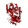
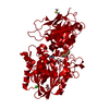
| Movie |
Movie viewer |
|---|---|
| Structure viewer | Molecule: MolmilJmol/JSmol |
- Downloads & links
-Download
| PDBx/mmCIF format | 7kc4.cif.gz | 154 KB | Display | PDBx/mmCIF format |
|---|---|---|---|---|
| PDB format | pdb7kc4.ent.gz | 126 KB | Display | PDB format |
| PDBx/mmJSON format | 7kc4.json.gz | Tree view | PDBx/mmJSON format | |
| Others |  Other downloads Other downloads |
-Validation report
| Arichive directory | https://data.pdbj.org/pub/pdb/validation_reports/kc/7kc4ftp://data.pdbj.org/pub/pdb/validation_reports/kc/7kc4 | HTTPS FTP |
|---|
-Related structure data
| Related structure data | 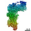 22806MC M: map data used to model this data C: citing same article ( |
|---|---|
| Similar structure data | |
| EM raw data | EMPIAR-10699 (Title: Human WLS in complex with WNT8A / Data size: 1.9 TB Data #1: Unaligned multi frame micrographs of WNT8A WLS complex [micrographs - multiframe]) |


-Links
 PDBj
PDBj


- Assembly
| Deposited unit | 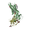
|
|---|---|
| 1 |
|
-Components
-Protein , 2 types, 2 molecules DB
| #1: Protein | Mass: 40052.359 Da / Num. of mol.: 1 Source method: isolated from a genetically manipulated source Source: (gene. exp.) Homo sapiens (human) / Gene: WNT8A, WNT8D / Plasmid: pEG BacMam vector / Cell line (production host): FreeStyle HEK293-F Cells / Production host: Homo sapiens (human) / References: UniProt: Q9H1J5 |
|---|---|
| #2: Protein | Mass: 65649.430 Da / Num. of mol.: 1 Source method: isolated from a genetically manipulated source Source: (gene. exp.) Homo sapiens (human) / Gene: WLS, C1orf139, GPR177, UNQ85/PRO18667 / Plasmid: pEG BacMam vector / Cell line (production host): FreeStyle HEK293-F cells / Production host: Homo sapiens (human) / References: UniProt: Q5T9L3 |
-Sugars , 2 types, 2 molecules
| #3: Polysaccharide | 2-acetamido-2-deoxy-beta-D-glucopyranose-(1-4)-2-acetamido-2-deoxy-beta-D-glucopyranose / Mass: 424.401 Da / Num. of mol.: 1 Source method: isolated from a genetically manipulated source |
|---|---|
| #4: Polysaccharide | alpha-D-mannopyranose-(1-3)-alpha-D-mannopyranose-(1-3)-[alpha-D-mannopyranose-(1-6)]beta-D- ...alpha-D-mannopyranose-(1-3)-alpha-D-mannopyranose-(1-3)-[alpha-D-mannopyranose-(1-6)]beta-D-mannopyranose-(1-4)-2-acetamido-2-deoxy-beta-D-glucopyranose-(1-4)-2-acetamido-2-deoxy-beta-D-glucopyranose / Mass: 1072.964 Da / Num. of mol.: 1 Source method: isolated from a genetically manipulated source |
-Non-polymers , 3 types, 5 molecules 
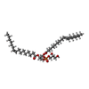

| #5: Chemical | ChemComp-PLM / Palmitic acid Mass: 256.424 Da / Num. of mol.: 1 / Source method: obtained synthetically / Formula: C16H32O2 / Feature type: SUBJECT OF INVESTIGATION Mass: 256.424 Da / Num. of mol.: 1 / Source method: obtained synthetically / Formula: C16H32O2 / Feature type: SUBJECT OF INVESTIGATION | ||
|---|---|---|---|
| #6: Chemical |  Mass: 746.991 Da / Num. of mol.: 3 / Source method: obtained synthetically / Formula: C40H75O10P Mass: 746.991 Da / Num. of mol.: 3 / Source method: obtained synthetically / Formula: C40H75O10P#7: Chemical | ChemComp-Y01 / |  Mass: 486.726 Da / Num. of mol.: 1 / Source method: obtained synthetically / Formula: C31H50O4 Mass: 486.726 Da / Num. of mol.: 1 / Source method: obtained synthetically / Formula: C31H50O4 |
-Details
| Has ligand of interest | Y |
|---|
-Experimental details
-Experiment
| Experiment | Method: ELECTRON MICROSCOPY |
|---|---|
| EM experiment | Aggregation state: PARTICLE / 3D reconstruction method: single particle reconstruction |
- Sample preparation
| Component | Name: Complex of WLS/Evi and WNT8A in nanodisc / Type: COMPLEX / Details: Nanodisc were formed using MSP1E3D1 and POPG lipid / Entity ID: #1-#2 / Source: RECOMBINANT | |||||||||||||||
|---|---|---|---|---|---|---|---|---|---|---|---|---|---|---|---|---|
| Molecular weight | Value: 0.1056 MDa / Experimental value: NO | |||||||||||||||
| Source (natural) | Organism: Homo sapiens (human) | |||||||||||||||
| Source (recombinant) | Organism: Homo sapiens (human) / Strain: FreeStyle 293-F Cells | |||||||||||||||
| Buffer solution | pH: 8 | |||||||||||||||
| Buffer component |
| |||||||||||||||
| Specimen | Conc.: 1 mg/ml / Embedding applied: NO / Shadowing applied: NO / Staining applied: NO / Vitrification applied: YES | |||||||||||||||
| Specimen support | Grid material: GOLD / Grid type: Quantifoil R0.6/1 | |||||||||||||||
| Vitrification | Instrument: FEI VITROBOT MARK IV / Cryogen name: ETHANE / Humidity: 95 % / Chamber temperature: 277 K |
- Electron microscopy imaging
| Microscopy | Model: FEI TITAN |
|---|---|
| Electron gun | Electron source: FIELD EMISSION GUN / Accelerating voltage: 300 kV / Illumination mode: FLOOD BEAM |
| Electron lens | Mode: BRIGHT FIELDBright-field microscopy |
| Image recording | Average exposure time: 3 sec. / Electron dose: 58 e/Å2 / Film or detector model: GATAN K3 BIOQUANTUM (6k x 4k) / Num. of grids imaged: 1 / Num. of real images: 12747 |
| Image scans | Sampling size: 5 µm / Width: 5760 / Height: 4092 |
- Processing
| Software | Name: PHENIX / Version: 1.19rc3_4024: / Classification: refinement | |||||||||||||||||||||||||
|---|---|---|---|---|---|---|---|---|---|---|---|---|---|---|---|---|---|---|---|---|---|---|---|---|---|---|
| EM software |
| |||||||||||||||||||||||||
| CTF correction | Details: Patch CTF / Type: PHASE FLIPPING AND AMPLITUDE CORRECTION | |||||||||||||||||||||||||
| Particle selection | Num. of particles selected: 4415933 | |||||||||||||||||||||||||
| 3D reconstruction | Resolution: 3.19 Å / Resolution method: FSC 0.143 CUT-OFF / Num. of particles: 27288 / Num. of class averages: 1 / Symmetry type: POINT | |||||||||||||||||||||||||
| Atomic model building | PDB-ID: 4F0A | |||||||||||||||||||||||||
| Refine LS restraints |
|