 Movie
Movie Controller

[English] 日本語
 Yorodumi
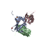
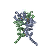
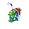
- PDB-7k31: Crystal structure of Endonuclease Q complex with 27-mer duplex su... -
+ Open data

- Basic information
| Entry | Database: PDB / ID: 7k31 | ||||||
|---|---|---|---|---|---|---|---|
| Title | Crystal structure of Endonuclease Q complex with 27-mer duplex substrate with dI at the active site | ||||||
 Components Components |
| ||||||
 Keywords Keywords | HYDROLASE/DNA /  HYDROLASE / deamination / HYDROLASE-DNA complex HYDROLASE / deamination / HYDROLASE-DNA complex | ||||||
| Function / homology | Conserved hypothetical protein CHP00375 / Polymerase/histidinol phosphatase-like / Rubredoxin, iron-binding site / Rubredoxin signature. / metal ion binding / DNA / DNA (> 10) / Phosphotransferase Function and homology information Function and homology information | ||||||
| Biological species |   Pyrococcus furiosus (archaea) Pyrococcus furiosus (archaea)synthetic construct (others) | ||||||
| Method | X-RAY DIFFRACTION / SYNCHROTRON / MOLECULAR REPLACEMENT / Resolution: 2.88 Å | ||||||
 Authors Authors | Shi, K. / Moeller, N.M. / Banerjee, S. / Yin, L. / Orellana, K. / Aihara, H. | ||||||
| Funding support |  United States, 1items United States, 1items
| ||||||
 Citation Citation | Journal: Proc.Natl.Acad.Sci.USA / Year: 2021 Title: Structural basis for recognition of distinct deaminated DNA lesions by endonuclease Q. Authors: Shi, K. / Moeller, N.H. / Banerjee, S. / McCann, J.L. / Carpenter, M.A. / Yin, L. / Moorthy, R. / Orellana, K. / Harki, D.A. / Harris, R.S. / Aihara, H. | ||||||
| History |
|
- Structure visualization
| Structure viewer | Molecule: MolmilJmol/JSmol |
|---|
- Downloads & links
-Download
| PDBx/mmCIF format | 7k31.cif.gz | 232.5 KB | Display | PDBx/mmCIF format |
|---|---|---|---|---|
| PDB format | pdb7k31.ent.gz | 181.5 KB | Display | PDB format |
| PDBx/mmJSON format | 7k31.json.gz | Tree view | PDBx/mmJSON format | |
| Others |  Other downloads Other downloads |
-Validation report
| Arichive directory | https://data.pdbj.org/pub/pdb/validation_reports/k3/7k31ftp://data.pdbj.org/pub/pdb/validation_reports/k3/7k31 | HTTPS FTP |
|---|
-Related structure data
| Related structure data |  7k30C  7k32C  7k33C  5zb8S S: Starting model for refinement C: citing same article ( |
|---|---|
| Similar structure data |


-Links
 PDBj
PDBj


- Assembly
| Deposited unit | 
| ||||||||||
|---|---|---|---|---|---|---|---|---|---|---|---|
| 1 |
| ||||||||||
| Unit cell |
|
-Components
-Protein , 1 types, 1 molecules A
| #1: Protein | Mass: 44456.328 Da / Num. of mol.: 1 Source method: isolated from a genetically manipulated source Source: (gene. exp.) Pyrococcus furiosus (archaea) / Production host:  Escherichia coli (E. coli) / References: UniProt: I6V2I0 Escherichia coli (E. coli) / References: UniProt: I6V2I0 |
|---|
-DNA chain , 2 types, 2 molecules BC
| #2: DNA chain | Mass: 8266.292 Da / Num. of mol.: 1 / Source method: obtained synthetically / Source: (synth.) synthetic construct (others) |
|---|---|
| #3: DNA chain | Mass: 8330.375 Da / Num. of mol.: 1 / Source method: obtained synthetically / Source: (synth.) synthetic construct (others) |
-Non-polymers , 5 types, 15 molecules 


| #4: Chemical | ChemComp-EDO / Ethylene glycol Mass: 62.068 Da / Num. of mol.: 1 / Source method: obtained synthetically / Formula: C2H6O2 Mass: 62.068 Da / Num. of mol.: 1 / Source method: obtained synthetically / Formula: C2H6O2 | ||||||
|---|---|---|---|---|---|---|---|
| #5: Chemical |  Mass: 65.409 Da / Num. of mol.: 3 / Source method: obtained synthetically / Formula: Zn Mass: 65.409 Da / Num. of mol.: 3 / Source method: obtained synthetically / Formula: Zn#6: Chemical | ChemComp-MG / |  Mass: 24.305 Da / Num. of mol.: 1 / Source method: isolated from a natural source / Formula: Mg Mass: 24.305 Da / Num. of mol.: 1 / Source method: isolated from a natural source / Formula: Mg#7: Chemical | Chloride Mass: 35.453 Da / Num. of mol.: 2 / Source method: isolated from a natural source / Formula: Cl Mass: 35.453 Da / Num. of mol.: 2 / Source method: isolated from a natural source / Formula: Cl#8: Water | ChemComp-HOH / | WaterMass: 18.015 Da / Num. of mol.: 8 / Source method: isolated from a natural source / Formula: H2O |
-Details
| Has ligand of interest | N |
|---|
-Experimental details
-Experiment
| Experiment | Method: X-RAY DIFFRACTION / Number of used crystals: 1 |
|---|
- Sample preparation
| Crystal | Density Matthews: 4.2 Å3/Da / Density % sol: 70.7 % |
|---|---|
| Crystal grow | Temperature: 295 K / Method: vapor diffusion, hanging drop / pH: 7.4 Details: 15 mM Tris-HCl pH 7.4, 0.15 M NaCl, 1 mM MgCl2, and 4 mM beta-mercaptoethanol |
-Data collection
| Diffraction | Mean temperature: 100 K / Serial crystal experiment: N | ||||||||||||||||||||||||||||||
|---|---|---|---|---|---|---|---|---|---|---|---|---|---|---|---|---|---|---|---|---|---|---|---|---|---|---|---|---|---|---|---|
| Diffraction source | Source: SYNCHROTRON / Site: APS / Beamline: 24-ID-C / Wavelength: 0.979 Å | ||||||||||||||||||||||||||||||
| Detector | Type: DECTRIS PILATUS3 S 6M / Detector: PIXEL / Date: Oct 13, 2019 | ||||||||||||||||||||||||||||||
| Radiation | Protocol: SINGLE WAVELENGTH / Monochromatic (M) / Laue (L): M / Scattering type: x-ray | ||||||||||||||||||||||||||||||
| Radiation wavelength | Wavelength: 0.979 Å / Relative weight: 1 | ||||||||||||||||||||||||||||||
| Reflection | Resolution: 2.85→87.47 Å / Num. obs: 22027 / % possible obs: 94.2 % / Redundancy: 2.3 % / Biso Wilson estimate: 42.82 Å2 / CC1/2: 0.99 / Rmerge(I) obs: 0.055 / Rpim(I) all: 0.045 / Rrim(I) all: 0.072 / Net I/σ(I): 10 | ||||||||||||||||||||||||||||||
| Reflection shell | Diffraction-ID: 1
|
- Processing
| Software |
| ||||||||||||||||||||||||||||||||||||||||||||||||||||||||||||||||||||||||
|---|---|---|---|---|---|---|---|---|---|---|---|---|---|---|---|---|---|---|---|---|---|---|---|---|---|---|---|---|---|---|---|---|---|---|---|---|---|---|---|---|---|---|---|---|---|---|---|---|---|---|---|---|---|---|---|---|---|---|---|---|---|---|---|---|---|---|---|---|---|---|---|---|---|
| Refinement | Method to determine structure: MOLECULAR REPLACEMENT Starting model: 5ZB8 Resolution: 2.88→87.47 Å / SU ML: 0.39 / Cross valid method: THROUGHOUT / σ(F): 1.98 / Phase error: 24.67 / Stereochemistry target values: ML
| ||||||||||||||||||||||||||||||||||||||||||||||||||||||||||||||||||||||||
| Solvent computation | Shrinkage radii: 0.9 Å / VDW probe radii: 1.11 Å / Solvent model: FLAT BULK SOLVENT MODEL | ||||||||||||||||||||||||||||||||||||||||||||||||||||||||||||||||||||||||
| Displacement parameters | Biso max: 157.65 Å2 / Biso mean: 58.4722 Å2 / Biso min: 4.9 Å2 | ||||||||||||||||||||||||||||||||||||||||||||||||||||||||||||||||||||||||
| Refinement step | Cycle: final / Resolution: 2.88→87.47 Å
| ||||||||||||||||||||||||||||||||||||||||||||||||||||||||||||||||||||||||
| LS refinement shell | Refine-ID: X-RAY DIFFRACTION / Rfactor Rfree error: 0 / Total num. of bins used: 6
| ||||||||||||||||||||||||||||||||||||||||||||||||||||||||||||||||||||||||
| Refinement TLS params. | L11: 0 °2 / L12: 0 °2 / L13: 0 °2 / L22: 0 °2 / L23: 0 °2 / L33: 0 °2 / S11: 0 Å ° / S12: 0 Å ° / S13: 0 Å ° / S21: 0 Å ° / S22: 0 Å ° / S23: 0 Å ° / S31: 0 Å ° / S32: 0 Å ° / S33: 0 Å ° / T11: 0 Å2 / T12: 0 Å2 / T13: 0 Å2 / T22: 0 Å2 / T23: 0 Å2 / T33: 0 Å2 / Method: refined / Refine-ID: X-RAY DIFFRACTION
| ||||||||||||||||||||||||||||||||||||||||||||||||||||||||||||||||||||||||
| Refinement TLS group |
|
