 Movie
Movie Controller


+ Open data

- Basic information
| Entry | Database: PDB / ID: 7k1w | ||||||
|---|---|---|---|---|---|---|---|
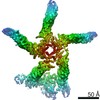
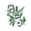
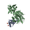
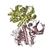
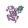
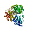
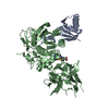
| Title | PIKfyve/Fig4/Vac14 complex centered on Fig4 - map3 | ||||||
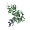
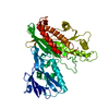
 Components Components | Fig4 Sac homology model | ||||||
 Keywords Keywords | LIPID BINDING PROTEIN /  Lipid kinase / Lipid phosphatase / protein complex Lipid kinase / Lipid phosphatase / protein complex | ||||||
| Function / homology |  Function and homology information Function and homology informationphosphatidylinositol-3,5-bisphosphate 5-phosphatase activity / Synthesis of PIPs at the late endosome membrane / Synthesis of PIPs at the early endosome membrane / Synthesis of PIPs at the Golgi membrane / phosphatidylinositol dephosphorylation / Hydrolases; Acting on ester bonds; Phosphoric-monoester hydrolases / phosphatidylinositol biosynthetic process / lipid droplet / late endosome membrane / early endosome membrane ...phosphatidylinositol-3,5-bisphosphate 5-phosphatase activity / Synthesis of PIPs at the late endosome membrane / Synthesis of PIPs at the early endosome membrane / Synthesis of PIPs at the Golgi membrane / phosphatidylinositol dephosphorylation / Hydrolases; Acting on ester bonds; Phosphoric-monoester hydrolases / phosphatidylinositol biosynthetic process / lipid droplet / late endosome membrane / early endosome membrane / endosome membrane / Golgi membrane / intracellular membrane-bounded organelleSimilarity search - Function | ||||||
| Biological species |  Homo sapiens (human) Homo sapiens (human) | ||||||
| Method | ELECTRON MICROSCOPY / single particle reconstruction / cryo EM / Resolution: 5.1 Å | ||||||
 Authors Authors | Lees, J.A. / Reinisch, K.M. / Li, P. | ||||||
| Funding support |  United States, 1items United States, 1items
| ||||||
 Citation Citation | Journal: Mol Cell / Year: 2020 Title: Insights into Lysosomal PI(3,5)P Homeostasis from a Structural-Biochemical Analysis of the PIKfyve Lipid Kinase Complex. Authors: Joshua A Lees / PeiQi Li / Nikit Kumar / Lois S Weisman / Karin M Reinisch / Abstract: The phosphoinositide PI(3,5)P, generated exclusively by the PIKfyve lipid kinase complex, is key for lysosomal biology. Here, we explore how PI(3,5)P levels within cells are regulated. We find the ...The phosphoinositide PI(3,5)P, generated exclusively by the PIKfyve lipid kinase complex, is key for lysosomal biology. Here, we explore how PI(3,5)P levels within cells are regulated. We find the PIKfyve complex comprises five copies of the scaffolding protein Vac14 and one copy each of the lipid kinase PIKfyve, generating PI(3,5)P from PI3P and the lipid phosphatase Fig4, reversing the reaction. Fig4 is active as a lipid phosphatase in the ternary complex, whereas PIKfyve within the complex cannot access membrane-incorporated phosphoinositides due to steric constraints. We find further that the phosphoinositide-directed activities of both PIKfyve and Fig4 are regulated by protein-directed activities within the complex. PIKfyve autophosphorylation represses its lipid kinase activity and stimulates Fig4 lipid phosphatase activity. Further, Fig4 is also a protein phosphatase acting on PIKfyve to stimulate its lipid kinase activity, explaining why catalytically active Fig4 is required for maximal PI(3,5)P production by PIKfyve in vivo. | ||||||
| History |
|
- Structure visualization
| Movie |
Movie viewer |
|---|---|
| Structure viewer | Molecule: MolmilJmol/JSmol |
- Downloads & links
-Download
| PDBx/mmCIF format | 7k1w.cif.gz | 104.9 KB | Display | PDBx/mmCIF format |
|---|---|---|---|---|
| PDB format | pdb7k1w.ent.gz | 74.2 KB | Display | PDB format |
| PDBx/mmJSON format | 7k1w.json.gz | Tree view | PDBx/mmJSON format | |
| Others |  Other downloads Other downloads |
-Validation report
| Arichive directory | https://data.pdbj.org/pub/pdb/validation_reports/k1/7k1wftp://data.pdbj.org/pub/pdb/validation_reports/k1/7k1w | HTTPS FTP |
|---|
-Related structure data
| Related structure data |  22631MC 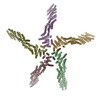 7k1yC  7k2vC M: map data used to model this data C: citing same article ( |
|---|---|
| Similar structure data |


-Links
 PDBj
PDBj- Assembly
| Deposited unit | 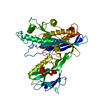
|
|---|---|
| 1 |
|
-Components
| #1: Protein | Mass: 105144.289 Da / Num. of mol.: 1 Source method: isolated from a genetically manipulated source Source: (gene. exp.) Homo sapiens (human) / Cell line (production host): Expi293F / Production host: Homo sapiens (human) / References: UniProt: Q92562*PLUS |
|---|
-Experimental details
-Experiment
| Experiment | Method: ELECTRON MICROSCOPY |
|---|---|
| EM experiment | Aggregation state: PARTICLE / 3D reconstruction method: single particle reconstruction |
- Sample preparation
| Component | Name: PIKfyve/Fig4/Vac14 complex / Type: COMPLEX / Entity ID: all / Source: RECOMBINANT |
|---|---|
| Molecular weight | Value: 4.28 MDa / Experimental value: NO |
| Source (natural) | Organism: Homo sapiens (human) |
| Source (recombinant) | Organism: Homo sapiens (human) / Cell: Expi293F |
| Buffer solution | pH: 7.8 |
| Specimen | Embedding applied: NO / Shadowing applied: NO / Staining applied: NO / Vitrification applied: YES |
| Vitrification | Cryogen name: ETHANE |
- Electron microscopy imaging
| Experimental equipment |  Model: Titan Krios / Image courtesy: FEI Company |
|---|---|
| Microscopy | Model: FEI TITAN KRIOS |
| Electron gun | Electron source: FIELD EMISSION GUN / Accelerating voltage: 300 kV / Illumination mode: FLOOD BEAM |
| Electron lens | Mode: BRIGHT FIELDBright-field microscopy / Cs: 2.7 mm |
| Image recording | Electron dose: 58.4 e/Å2 / Film or detector model: GATAN K2 SUMMIT (4k x 4k) |
- Processing
| Software | Name: PHENIX / Version: 1.16_3549: / Classification: refinement | ||||||||||||||||||||||||
|---|---|---|---|---|---|---|---|---|---|---|---|---|---|---|---|---|---|---|---|---|---|---|---|---|---|
| CTF correction | Type: NONE | ||||||||||||||||||||||||
| 3D reconstruction | Resolution: 5.1 Å / Resolution method: FSC 0.143 CUT-OFF / Num. of particles: 19998 / Symmetry type: POINT | ||||||||||||||||||||||||
| Refine LS restraints |
|
