 Movie
Movie Controller

[English] 日本語
 Yorodumi
- PDB-7jvb: Crystal structure of the SARS-CoV-2 spike receptor-binding domain... -
+ Open data

- Basic information
| Entry | Database: PDB / ID: 7jvb | ||||||
|---|---|---|---|---|---|---|---|
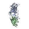
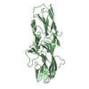
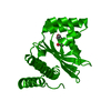
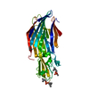
| Title | Crystal structure of the SARS-CoV-2 spike receptor-binding domain (RBD) with nanobody Nb20 | ||||||
 Components Components |
| ||||||
 Keywords Keywords |  VIRAL PROTEIN / SARS-CoV-2 / COVID-19 / nanobody / spike protein / receptor-binding domain VIRAL PROTEIN / SARS-CoV-2 / COVID-19 / nanobody / spike protein / receptor-binding domain | ||||||
| Function / homology |  Function and homology information Function and homology informationMaturation of spike protein / viral translation / Translation of Structural Proteins / Virion Assembly and Release / host cell surface / host extracellular space / suppression by virus of host tetherin activity / Induction of Cell-Cell Fusion / structural constituent of virion / host cell endoplasmic reticulum-Golgi intermediate compartment membrane ...Maturation of spike protein / viral translation / Translation of Structural Proteins / Virion Assembly and Release / host cell surface / host extracellular space / suppression by virus of host tetherin activity / Induction of Cell-Cell Fusion / structural constituent of virion / host cell endoplasmic reticulum-Golgi intermediate compartment membrane / entry receptor-mediated virion attachment to host cell / receptor-mediated endocytosis of virus by host cell / Attachment and Entry / membrane fusion / positive regulation of viral entry into host cell / receptor-mediated virion attachment to host cell / receptor ligand activity / host cell surface receptor binding / fusion of virus membrane with host plasma membrane / fusion of virus membrane with host endosome membrane / viral envelope / virion attachment to host cell / SARS-CoV-2 activates/modulates innate and adaptive immune responses / host cell plasma membrane / virion membrane / membrane / identical protein binding / plasma membraneSimilarity search - Function | ||||||
| Biological species |   Severe acute respiratory syndrome coronavirus 2 Severe acute respiratory syndrome coronavirus 2 Lama glama (llama) Lama glama (llama) | ||||||
| Method | X-RAY DIFFRACTION / SYNCHROTRON / MOLECULAR REPLACEMENT / Resolution: 3.287 Å | ||||||
 Authors Authors | Xiang, Y. / Xiao, Z. / Liu, H. / Sang, Z. / Schneidman-Duhovny, D. / Zhang, C. / Shi, Y. | ||||||
| Funding support |  United States, 1items United States, 1items
| ||||||
 Citation Citation | Journal: Science / Year: 2020 Title: Versatile and multivalent nanobodies efficiently neutralize SARS-CoV-2. Authors: Xiang, Y. / Nambulli, S. / Xiao, Z. / Liu, H. / Sang, Z. / Duprex, W.P. / Schneidman-Duhovny, D. / Zhang, C. / Shi, Y. | ||||||
| History |
|
- Structure visualization
| Structure viewer | Molecule: MolmilJmol/JSmol |
|---|
- Downloads & links
-Download
| PDBx/mmCIF format | 7jvb.cif.gz | 257.8 KB | Display | PDBx/mmCIF format |
|---|---|---|---|---|
| PDB format | pdb7jvb.ent.gz | 208.5 KB | Display | PDB format |
| PDBx/mmJSON format | 7jvb.json.gz | Tree view | PDBx/mmJSON format | |
| Others |  Other downloads Other downloads |
-Validation report
| Arichive directory | https://data.pdbj.org/pub/pdb/validation_reports/jv/7jvbftp://data.pdbj.org/pub/pdb/validation_reports/jv/7jvb | HTTPS FTP |
|---|
-Related structure data
| Related structure data | 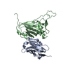 6waqS S: Starting model for refinement |
|---|---|
| Similar structure data |


-Links
 PDBj
PDBj


- Assembly
| Deposited unit | 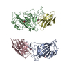
| ||||||||||
|---|---|---|---|---|---|---|---|---|---|---|---|
| 1 | 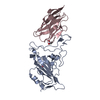
| ||||||||||
| 2 | 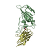
| ||||||||||
| Unit cell |
|
-Components
| #1: Protein | Mass: 25179.389 Da / Num. of mol.: 2 Source method: isolated from a genetically manipulated source Source: (gene. exp.) Severe acute respiratory syndrome coronavirus 2Gene: S, 2 / Production host:   Spodoptera frugiperda (fall armyworm) / References: UniProt: P0DTC2 Spodoptera frugiperda (fall armyworm) / References: UniProt: P0DTC2#2: Antibody | Mass: 12553.048 Da / Num. of mol.: 2 Source method: isolated from a genetically manipulated source Source: (gene. exp.) Lama glama (llama) / Production host:  Escherichia coli BL21 (bacteria) Escherichia coli BL21 (bacteria)#3: Chemical | ChemComp-CAC / Cacodylic acid  Mass: 136.989 Da / Num. of mol.: 4 / Source method: obtained synthetically / Formula: C2H6AsO2 Mass: 136.989 Da / Num. of mol.: 4 / Source method: obtained synthetically / Formula: C2H6AsO2Has ligand of interest | N | |
|---|
-Experimental details
-Experiment
| Experiment | Method: X-RAY DIFFRACTION / Number of used crystals: 1 |
|---|
- Sample preparation
| Crystal | Density Matthews: 3.6 Å3/Da / Density % sol: 65.87 % |
|---|---|
| Crystal grow | Temperature: 290 K / Method: vapor diffusion, sitting drop / pH: 6.5 Details: 100 mM sodium cacodylate pH 6.5, 1 M sodium citrate |
-Data collection
| Diffraction | Mean temperature: 80 K / Ambient temp details: liquid nitrogen / Serial crystal experiment: N |
|---|---|
| Diffraction source | Source: SYNCHROTRON / Site: APS / Beamline: 23-ID-B / Wavelength: 1 Å |
| Detector | Type: DECTRIS EIGER X 16M / Detector: PIXEL / Date: Aug 7, 2020 |
| Radiation | Protocol: SINGLE WAVELENGTH / Monochromatic (M) / Laue (L): M / Scattering type: x-ray |
| Radiation wavelength | Wavelength: 1 Å / Relative weight: 1 |
| Reflection | Resolution: 3.287→38.96 Å / Num. obs: 17442 / % possible obs: 96.34 % / Redundancy: 6.2 % / Biso Wilson estimate: 98.45 Å2 / CC1/2: 1 / Rmerge(I) obs: 0.156 / Rpim(I) all: 0.108 / Net I/σ(I): 7.8 |
| Reflection shell | Resolution: 3.287→3.405 Å / Rmerge(I) obs: 0.591 / Num. unique obs: 1391 / CC1/2: 0.811 / Rpim(I) all: 0.445 |
- Processing
| Software |
| |||||||||||||||||||||||||||||||||||||||||||||||||
|---|---|---|---|---|---|---|---|---|---|---|---|---|---|---|---|---|---|---|---|---|---|---|---|---|---|---|---|---|---|---|---|---|---|---|---|---|---|---|---|---|---|---|---|---|---|---|---|---|---|---|
| Refinement | Method to determine structure: MOLECULAR REPLACEMENT Starting model: 6WAQ Resolution: 3.287→38.959 Å / SU ML: 0.4 / Cross valid method: FREE R-VALUE / σ(F): 1.34 / Phase error: 37.56 / Stereochemistry target values: ML
| |||||||||||||||||||||||||||||||||||||||||||||||||
| Solvent computation | Shrinkage radii: 0.9 Å / VDW probe radii: 1.11 Å / Solvent model: FLAT BULK SOLVENT MODEL | |||||||||||||||||||||||||||||||||||||||||||||||||
| Refinement step | Cycle: LAST / Resolution: 3.287→38.959 Å
| |||||||||||||||||||||||||||||||||||||||||||||||||
| Refine LS restraints |
| |||||||||||||||||||||||||||||||||||||||||||||||||
| LS refinement shell |
| |||||||||||||||||||||||||||||||||||||||||||||||||
| Refinement TLS params. | Method: refined / Origin x: -16.1778 Å / Origin y: -15.3348 Å / Origin z: -27.1171 Å
| |||||||||||||||||||||||||||||||||||||||||||||||||
| Refinement TLS group | Selection details: all |
