 Movie
Movie Controller


+ Open data

- Basic information
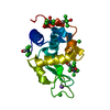
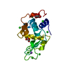
| Entry | Database: PDB / ID: 7jmu | ||||||
|---|---|---|---|---|---|---|---|
| Title | Hen egg-white lysozyme with ionic liquid ethylammonium nitrate | ||||||
 Components Components | Lysozyme C | ||||||
 Keywords Keywords |  HYDROLASE / lysozyme / ionic liquid / nitrate HYDROLASE / lysozyme / ionic liquid / nitrate | ||||||
| Function / homology |  Function and homology information Function and homology informationLactose synthesis / Antimicrobial peptides / Neutrophil degranulation / beta-N-acetylglucosaminidase activity / cell wall macromolecule catabolic process / lysozyme / lysozyme activity / killing of cells of another organism / defense response to Gram-negative bacterium / defense response to Gram-positive bacterium ...Lactose synthesis / Antimicrobial peptides / Neutrophil degranulation / beta-N-acetylglucosaminidase activity / cell wall macromolecule catabolic process / lysozyme / lysozyme activity / killing of cells of another organism / defense response to Gram-negative bacterium / defense response to Gram-positive bacterium / defense response to bacterium / endoplasmic reticulum / extracellular space / identical protein binding / cytoplasmSimilarity search - Function | ||||||
| Biological species |  Gallus gallus (chicken) Gallus gallus (chicken) | ||||||
| Method | X-RAY DIFFRACTION / SYNCHROTRON / MOLECULAR REPLACEMENT / Resolution: 1.2 Å | ||||||
 Authors Authors | Han, Q. / Darmanin, C. / Smith, K. / Greaves, T. / Drummond, C. | ||||||
 Citation Citation | Journal: J Colloid Interface Sci / Year: 2021 Title: Lysozyme conformational changes with ionic liquids: Spectroscopic, small angle x-ray scattering and crystallographic study. Authors: Han, Q. / Smith, K.M. / Darmanin, C. / Ryan, T.M. / Drummond, C.J. / Greaves, T.L. | ||||||
| History |
|
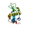
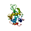
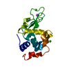
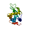
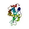
- Structure visualization
| Structure viewer | Molecule: MolmilJmol/JSmol |
|---|
- Downloads & links
-Download
| PDBx/mmCIF format | 7jmu.cif.gz | 212.5 KB | Display | PDBx/mmCIF format |
|---|---|---|---|---|
| PDB format | pdb7jmu.ent.gz | 171.1 KB | Display | PDB format |
| PDBx/mmJSON format | 7jmu.json.gz | Tree view | PDBx/mmJSON format | |
| Others |  Other downloads Other downloads |
-Validation report
| Arichive directory | https://data.pdbj.org/pub/pdb/validation_reports/jm/7jmuftp://data.pdbj.org/pub/pdb/validation_reports/jm/7jmu | HTTPS FTP |
|---|
-Related structure data
| Related structure data | 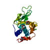 1dpwS S: Starting model for refinement |
|---|---|
| Similar structure data | |
| Experimental dataset #1 | Data reference: 10.25439/rmt.12705056 / Data set type: small-angle scattering data / Metadata reference: 10.25439/rmt.12705056 |


-Links
 PDBj
PDBj


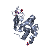
- Assembly
| Deposited unit | 
| ||||||||
|---|---|---|---|---|---|---|---|---|---|
| 1 | 
| ||||||||
| 2 | 
| ||||||||
| Unit cell |
|
-Components
| #1: Protein | Mass: 14331.160 Da / Num. of mol.: 2 / Source method: isolated from a natural source / Source: (natural) Gallus gallus (chicken) / Tissue: egg white / References: UniProt: P00698, lysozyme#2: Chemical | ChemComp-NO3 / Nitrate  Mass: 62.005 Da / Num. of mol.: 15 / Source method: obtained synthetically / Formula: NO3 / Feature type: SUBJECT OF INVESTIGATION Mass: 62.005 Da / Num. of mol.: 15 / Source method: obtained synthetically / Formula: NO3 / Feature type: SUBJECT OF INVESTIGATION#3: Water | ChemComp-HOH / | Water Mass: 18.015 Da / Num. of mol.: 252 / Source method: isolated from a natural source / Formula: H2O Mass: 18.015 Da / Num. of mol.: 252 / Source method: isolated from a natural source / Formula: H2OHas ligand of interest | Y | |
|---|
-Experimental details
-Experiment
| Experiment | Method: X-RAY DIFFRACTION / Number of used crystals: 1 |
|---|
- Sample preparation
| Crystal | Density Matthews: 1.8 Å3/Da / Density % sol: 31.58 % |
|---|---|
| Crystal grow | Temperature: 298.15 K / Method: batch mode / pH: 8 / Details: 5.7% w/v ethylammonium nitrate |
-Data collection
| Diffraction | Mean temperature: 100 K / Serial crystal experiment: N | ||||||||||||||||||||||||||||||
|---|---|---|---|---|---|---|---|---|---|---|---|---|---|---|---|---|---|---|---|---|---|---|---|---|---|---|---|---|---|---|---|
| Diffraction source | Source: SYNCHROTRON / Site: Australian Synchrotron  / Beamline: MX2 / Wavelength: 0.953646 Å / Beamline: MX2 / Wavelength: 0.953646 Å | ||||||||||||||||||||||||||||||
| Detector | Type: DECTRIS EIGER X 16M / Detector: PIXEL / Date: Oct 16, 2019 | ||||||||||||||||||||||||||||||
| Radiation | Protocol: SINGLE WAVELENGTH / Monochromatic (M) / Laue (L): M / Scattering type: x-ray | ||||||||||||||||||||||||||||||
| Radiation wavelength | Wavelength: 0.953646 Å / Relative weight: 1 | ||||||||||||||||||||||||||||||
| Reflection | Resolution: 1.2→43.19 Å / Num. obs: 60488 / % possible obs: 95.5 % / Redundancy: 6.9 % / CC1/2: 0.999 / Rmerge(I) obs: 0.046 / Rpim(I) all: 0.019 / Rrim(I) all: 0.049 / Net I/σ(I): 19.5 / Num. measured all: 417805 / Scaling rejects: 60 | ||||||||||||||||||||||||||||||
| Reflection shell | Diffraction-ID: 1
|
- Processing
| Software |
| |||||||||||||||||||||||||||||||||||||||||||||||||||||||||||||||||
|---|---|---|---|---|---|---|---|---|---|---|---|---|---|---|---|---|---|---|---|---|---|---|---|---|---|---|---|---|---|---|---|---|---|---|---|---|---|---|---|---|---|---|---|---|---|---|---|---|---|---|---|---|---|---|---|---|---|---|---|---|---|---|---|---|---|---|
| Refinement | Method to determine structure: MOLECULAR REPLACEMENT Starting model: PDB entry 1DPW Resolution: 1.2→29.82 Å / Cor.coef. Fo:Fc: 0.976 / Cor.coef. Fo:Fc free: 0.966 / SU B: 1.543 / SU ML: 0.031 / Cross valid method: THROUGHOUT / σ(F): 0 / ESU R: 0.047 / ESU R Free: 0.046 / Stereochemistry target values: MAXIMUM LIKELIHOOD Details: HYDROGENS HAVE BEEN ADDED IN THE RIDING POSITIONS U VALUES : REFINED INDIVIDUALLY
| |||||||||||||||||||||||||||||||||||||||||||||||||||||||||||||||||
| Solvent computation | Ion probe radii: 0.8 Å / Shrinkage radii: 0.8 Å / VDW probe radii: 1.2 Å / Solvent model: MASK | |||||||||||||||||||||||||||||||||||||||||||||||||||||||||||||||||
| Displacement parameters | Biso max: 72.85 Å2 / Biso mean: 15.044 Å2 / Biso min: 6.37 Å2
| |||||||||||||||||||||||||||||||||||||||||||||||||||||||||||||||||
| Refinement step | Cycle: final / Resolution: 1.2→29.82 Å
| |||||||||||||||||||||||||||||||||||||||||||||||||||||||||||||||||
| Refine LS restraints |
| |||||||||||||||||||||||||||||||||||||||||||||||||||||||||||||||||
| LS refinement shell | Resolution: 1.2→1.231 Å / Rfactor Rfree error: 0 / Total num. of bins used: 20
|
