 Movie
Movie Controller

[English] 日本語
 Yorodumi
- PDB-7fw5: Crystal Structure of human FABP4 with active site mutated to that... -
+ Open data

- Basic information
| Entry | Database: PDB / ID: 7fw5 | ||||||
|---|---|---|---|---|---|---|---|
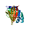
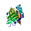
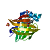
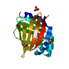
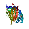
| Title | Crystal Structure of human FABP4 with active site mutated to that of FABP3 in complex with palmitate | ||||||
 Components Components | Fatty acid-binding protein, adipocyte | ||||||
 Keywords Keywords | LIPID BINDING PROTEIN /  FATTY ACID BINDING PROTEIN / CYTOPLASM / LIPID-BINDING / TRANSPORT / PROTEIN BINDING FATTY ACID BINDING PROTEIN / CYTOPLASM / LIPID-BINDING / TRANSPORT / PROTEIN BINDING | ||||||
| Function / homology |  Function and homology informationhormone receptor binding / long-chain fatty acid transmembrane transporter activity / long-chain fatty acid binding / cellular response to lithium ion / Triglyceride catabolism / white fat cell differentiation / fatty acid transport / long-chain fatty acid transport / brown fat cell differentiation / lipid droplet ...hormone receptor binding / long-chain fatty acid transmembrane transporter activity / long-chain fatty acid binding / cellular response to lithium ion / Triglyceride catabolism / white fat cell differentiation / fatty acid transport / long-chain fatty acid transport / brown fat cell differentiation / lipid droplet / cholesterol homeostasis / fatty acid binding / response to bacterium / positive regulation of inflammatory response / Transcriptional regulation of white adipocyte differentiation / cellular response to tumor necrosis factor / positive regulation of cold-induced thermogenesis / negative regulation of DNA-templated transcription / extracellular exosome / nucleus / cytosol / cytoplasm Function and homology informationhormone receptor binding / long-chain fatty acid transmembrane transporter activity / long-chain fatty acid binding / cellular response to lithium ion / Triglyceride catabolism / white fat cell differentiation / fatty acid transport / long-chain fatty acid transport / brown fat cell differentiation / lipid droplet ...hormone receptor binding / long-chain fatty acid transmembrane transporter activity / long-chain fatty acid binding / cellular response to lithium ion / Triglyceride catabolism / white fat cell differentiation / fatty acid transport / long-chain fatty acid transport / brown fat cell differentiation / lipid droplet / cholesterol homeostasis / fatty acid binding / response to bacterium / positive regulation of inflammatory response / Transcriptional regulation of white adipocyte differentiation / cellular response to tumor necrosis factor / positive regulation of cold-induced thermogenesis / negative regulation of DNA-templated transcription / extracellular exosome / nucleus / cytosol / cytoplasmSimilarity search - Function | ||||||
| Biological species |  Homo sapiens (human) Homo sapiens (human) | ||||||
| Method | X-RAY DIFFRACTION / SYNCHROTRON / MOLECULAR REPLACEMENT / Resolution: 1.15 Å | ||||||
 Authors Authors | Ehler, A. / Benz, J. / Obst, U. / Rudolph, M.G. | ||||||
| Funding support |  Switzerland, 1items Switzerland, 1items
| ||||||
 Citation Citation | Journal: To be published Title: Crystal Structure of a human FABP4 complex Authors: Obst, U. / Magnone, C. / Kuhn, B. / Rudolph, M.G. | ||||||
| History |
|
- Structure visualization
| Structure viewer | Molecule: MolmilJmol/JSmol |
|---|
- Downloads & links
-Download
| PDBx/mmCIF format | 7fw5.cif.gz | 77.8 KB | Display | PDBx/mmCIF format |
|---|---|---|---|---|
| PDB format | pdb7fw5.ent.gz | 57.5 KB | Display | PDB format |
| PDBx/mmJSON format | 7fw5.json.gz | Tree view | PDBx/mmJSON format | |
| Others |  Other downloads Other downloads |
-Validation report
| Arichive directory | https://data.pdbj.org/pub/pdb/validation_reports/fw/7fw5ftp://data.pdbj.org/pub/pdb/validation_reports/fw/7fw5 | HTTPS FTP |
|---|
-Group deposition
| ID | G_1002264 (222 entries) |
|---|---|
| Title | To be published |
| Type | undefined |
| Description | A set of fabp crystal structures |


-Related structure data
| Similar structure data |
|---|
-Links
 PDBj
PDBj


- Assembly
| Deposited unit | 
| ||||||||
|---|---|---|---|---|---|---|---|---|---|
| 1 |
| ||||||||
| Unit cell |
|
-Components
| #1: Protein | Mass: 15060.242 Da / Num. of mol.: 1 Source method: isolated from a genetically manipulated source Source: (gene. exp.) Homo sapiens (human) / Gene: FABP4 / Plasmid: PET15b / Production host:  Escherichia coli BL21(DE3) (bacteria) / References: UniProt: P15090 Escherichia coli BL21(DE3) (bacteria) / References: UniProt: P15090 |
|---|---|
| #2: Chemical | ChemComp-PLM / Palmitic acid  Mass: 256.424 Da / Num. of mol.: 1 / Source method: obtained synthetically / Formula: C16H32O2 Mass: 256.424 Da / Num. of mol.: 1 / Source method: obtained synthetically / Formula: C16H32O2 |
| #3: Water | ChemComp-HOH / Water Mass: 18.015 Da / Num. of mol.: 182 / Source method: isolated from a natural source / Formula: H2O Mass: 18.015 Da / Num. of mol.: 182 / Source method: isolated from a natural source / Formula: H2O |
-Experimental details
-Experiment
| Experiment | Method: X-RAY DIFFRACTION / Number of used crystals: 1 |
|---|
- Sample preparation
| Crystal | Density Matthews: 2.48 Å3/Da / Density % sol: 50.33 % |
|---|---|
| Crystal grow | Temperature: 293 K / Method: vapor diffusion, sitting drop / pH: 7 Details: protein in 25mM Tris/HCl pH 7.5 100mM NaCl, see also PMID 27658368 |
-Data collection
| Diffraction | Mean temperature: 100 K | ||||||||||||||||||||||||||||||||||||||||||||||||||||||||||||||||||||||||||||||||||||||||||||||||||||||||||||||||||||||||||||||||||||||||||||||||||||||||||||||||||||||||||||||||||||||||||||||||||||||||||||||||||
|---|---|---|---|---|---|---|---|---|---|---|---|---|---|---|---|---|---|---|---|---|---|---|---|---|---|---|---|---|---|---|---|---|---|---|---|---|---|---|---|---|---|---|---|---|---|---|---|---|---|---|---|---|---|---|---|---|---|---|---|---|---|---|---|---|---|---|---|---|---|---|---|---|---|---|---|---|---|---|---|---|---|---|---|---|---|---|---|---|---|---|---|---|---|---|---|---|---|---|---|---|---|---|---|---|---|---|---|---|---|---|---|---|---|---|---|---|---|---|---|---|---|---|---|---|---|---|---|---|---|---|---|---|---|---|---|---|---|---|---|---|---|---|---|---|---|---|---|---|---|---|---|---|---|---|---|---|---|---|---|---|---|---|---|---|---|---|---|---|---|---|---|---|---|---|---|---|---|---|---|---|---|---|---|---|---|---|---|---|---|---|---|---|---|---|---|---|---|---|---|---|---|---|---|---|---|---|---|---|---|---|---|
| Diffraction source | Source: SYNCHROTRON / Site: SLS / Beamline: X10SA / Wavelength: 1 Å | ||||||||||||||||||||||||||||||||||||||||||||||||||||||||||||||||||||||||||||||||||||||||||||||||||||||||||||||||||||||||||||||||||||||||||||||||||||||||||||||||||||||||||||||||||||||||||||||||||||||||||||||||||
| Detector | Type: PSI PILATUS 6M / Detector: PIXEL / Date: Jan 28, 2011 | ||||||||||||||||||||||||||||||||||||||||||||||||||||||||||||||||||||||||||||||||||||||||||||||||||||||||||||||||||||||||||||||||||||||||||||||||||||||||||||||||||||||||||||||||||||||||||||||||||||||||||||||||||
| Radiation | Protocol: SINGLE WAVELENGTH / Monochromatic (M) / Laue (L): M / Scattering type: x-ray | ||||||||||||||||||||||||||||||||||||||||||||||||||||||||||||||||||||||||||||||||||||||||||||||||||||||||||||||||||||||||||||||||||||||||||||||||||||||||||||||||||||||||||||||||||||||||||||||||||||||||||||||||||
| Radiation wavelength | Wavelength: 1 Å / Relative weight: 1 | ||||||||||||||||||||||||||||||||||||||||||||||||||||||||||||||||||||||||||||||||||||||||||||||||||||||||||||||||||||||||||||||||||||||||||||||||||||||||||||||||||||||||||||||||||||||||||||||||||||||||||||||||||
| Reflection | Resolution: 1.15→44.72 Å / Num. obs: 53727 / % possible obs: 99.5 % / Redundancy: 6.428 % / Biso Wilson estimate: 18.615 Å2 / CC1/2: 0.999 / Rmerge(I) obs: 0.048 / Rrim(I) all: 0.053 / Χ2: 0.878 / Net I/σ(I): 15.09 / Num. measured all: 345384 / Scaling rejects: 472 | ||||||||||||||||||||||||||||||||||||||||||||||||||||||||||||||||||||||||||||||||||||||||||||||||||||||||||||||||||||||||||||||||||||||||||||||||||||||||||||||||||||||||||||||||||||||||||||||||||||||||||||||||||
| Reflection shell | Diffraction-ID: 1
|
- Processing
| Software |
| |||||||||||||||||||||||||||||||||||||||||||||||||||||||||||||||||||||||||||
|---|---|---|---|---|---|---|---|---|---|---|---|---|---|---|---|---|---|---|---|---|---|---|---|---|---|---|---|---|---|---|---|---|---|---|---|---|---|---|---|---|---|---|---|---|---|---|---|---|---|---|---|---|---|---|---|---|---|---|---|---|---|---|---|---|---|---|---|---|---|---|---|---|---|---|---|---|
| Refinement | Method to determine structure: MOLECULAR REPLACEMENT Starting model: inhouse model Resolution: 1.15→44.72 Å / Cor.coef. Fo:Fc: 0.971 / Cor.coef. Fo:Fc free: 0.958 / SU B: 1.672 / SU ML: 0.034 / Cross valid method: THROUGHOUT / σ(F): 0 / ESU R: 0.042 / ESU R Free: 0.043 / Stereochemistry target values: MAXIMUM LIKELIHOOD Details: FABP43 is mutated FABP4 with ligand binding site from FABP3
| |||||||||||||||||||||||||||||||||||||||||||||||||||||||||||||||||||||||||||
| Solvent computation | Ion probe radii: 0.8 Å / Shrinkage radii: 0.8 Å / VDW probe radii: 1.2 Å / Solvent model: BABINET MODEL WITH MASK | |||||||||||||||||||||||||||||||||||||||||||||||||||||||||||||||||||||||||||
| Displacement parameters | Biso max: 55.61 Å2 / Biso mean: 15.244 Å2 / Biso min: 10.85 Å2
| |||||||||||||||||||||||||||||||||||||||||||||||||||||||||||||||||||||||||||
| Refinement step | Cycle: final / Resolution: 1.15→44.72 Å
| |||||||||||||||||||||||||||||||||||||||||||||||||||||||||||||||||||||||||||
| Refine LS restraints |
| |||||||||||||||||||||||||||||||||||||||||||||||||||||||||||||||||||||||||||
| LS refinement shell | Resolution: 1.15→1.18 Å / Rfactor Rfree error: 0 / Total num. of bins used: 20
|
