 Movie
Movie Controller


+ Open data

- Basic information
| Entry | Database: PDB / ID: 7fjr | ||||||
|---|---|---|---|---|---|---|---|
| Title | Structure of a mutant of OspA | ||||||
 Components Components | Outer surface protein A | ||||||
 Keywords Keywords |  LIPOPROTEIN / Outer surface protein A / OspA / LIPID BINDING PROTEIN LIPOPROTEIN / Outer surface protein A / OspA / LIPID BINDING PROTEIN | ||||||
| Function / homology | Outer surface lipoprotein, Borrelia / Outer surface lipoprotein domain superfamily / Borrelia lipoprotein / cell outer membrane / Prokaryotic membrane lipoprotein lipid attachment site profile. / cell surface / membrane / DI(HYDROXYETHYL)ETHER / Outer surface protein A Function and homology information Function and homology information | ||||||
| Biological species |  Borreliella burgdorferi (Lyme disease spirochete) Borreliella burgdorferi (Lyme disease spirochete) | ||||||
| Method | X-RAY DIFFRACTION / SYNCHROTRON / MOLECULAR REPLACEMENT / Resolution: 2.6 Å | ||||||
 Authors Authors | Shiga, S. / Makabe, K. | ||||||
| Funding support | 1items
| ||||||
 Citation Citation | Journal: J.Mol.Biol. / Year: 2024 Title: beta-Strand-mediated Domain-swapping in the Absence of Hydrophobic Core Repacking. Authors: Kiya, M. / Shiga, S. / Ding, P. / Koide, S. / Makabe, K. | ||||||
| History |
|
- Structure visualization
| Structure viewer | Molecule: MolmilJmol/JSmol |
|---|
- Downloads & links
-Download
| PDBx/mmCIF format | 7fjr.cif.gz | 122.8 KB | Display | PDBx/mmCIF format |
|---|---|---|---|---|
| PDB format | pdb7fjr.ent.gz | 79.7 KB | Display | PDB format |
| PDBx/mmJSON format | 7fjr.json.gz | Tree view | PDBx/mmJSON format | |
| Others |  Other downloads Other downloads |
-Validation report
| Arichive directory | https://data.pdbj.org/pub/pdb/validation_reports/fj/7fjrftp://data.pdbj.org/pub/pdb/validation_reports/fj/7fjr | HTTPS FTP |
|---|
-Related structure data
| Related structure data |  6kwjC 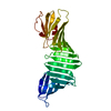 6kwuC  6kwvC  6ljyC 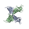 7fddC  2g8cS S: Starting model for refinement C: citing same article ( |
|---|---|
| Similar structure data |
-Links
 PDBj
PDBj


- Assembly
| Deposited unit | 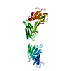
| ||||||||||||
|---|---|---|---|---|---|---|---|---|---|---|---|---|---|
| 1 | 
| ||||||||||||
| Unit cell |
|
-Components
| #1: Protein | Mass: 25609.604 Da / Num. of mol.: 1 Mutation: E37S, E45S, K46S, K48A, K60A, K64S, K83A, E104S, K107S, Deletion 117-119, insertion GG, Deletion 124-127, Deletion 130-133, insertion CTC, K239S, E240S, K254S Source method: isolated from a genetically manipulated source Source: (gene. exp.) Borreliella burgdorferi (Lyme disease spirochete)Gene: ospA, BB_A15 / Production host: Escherichia coli BL21(DE3) (bacteria) / Strain (production host): BL21(DE3) / References: UniProt: P0CL66 | ||||
|---|---|---|---|---|---|
| #2: Chemical | Diethylene glycol  Mass: 106.120 Da / Num. of mol.: 2 / Source method: obtained synthetically / Formula: C4H10O3 Mass: 106.120 Da / Num. of mol.: 2 / Source method: obtained synthetically / Formula: C4H10O3#3: Water | ChemComp-HOH / | Water Mass: 18.015 Da / Num. of mol.: 7 / Source method: isolated from a natural source / Formula: H2O Mass: 18.015 Da / Num. of mol.: 7 / Source method: isolated from a natural source / Formula: H2OHas ligand of interest | N | |
-Experimental details
-Experiment
| Experiment | Method: X-RAY DIFFRACTION / Number of used crystals: 1 |
|---|
- Sample preparation
| Crystal | Density Matthews: 3.78 Å3/Da / Density % sol: 67.5 % |
|---|---|
| Crystal grow | Temperature: 292 K / Method: vapor diffusion, hanging drop / pH: 8 Details: Protein: 16.0 mg/mL, 35% Polyethylene glycol 400, 0.1 M Tris-HCl |
-Data collection
| Diffraction | Mean temperature: 100 K / Serial crystal experiment: N |
|---|---|
| Diffraction source | Source: SYNCHROTRON / Site: Photon Factory  / Beamline: BL-5A / Wavelength: 1 Å / Beamline: BL-5A / Wavelength: 1 Å |
| Detector | Type: DECTRIS PILATUS3 S 6M / Detector: PIXEL / Date: May 12, 2021 |
| Radiation | Protocol: SINGLE WAVELENGTH / Monochromatic (M) / Laue (L): M / Scattering type: x-ray |
| Radiation wavelength | Wavelength: 1 Å / Relative weight: 1 |
| Reflection | Resolution: 2.6→19.69 Å / Num. obs: 12935 / % possible obs: 100 % / Redundancy: 17.7 % / Biso Wilson estimate: 53.83 Å2 / Rmerge(I) obs: 0.053 / Net I/σ(I): 49.3 |
| Reflection shell | Resolution: 2.6→2.69 Å / Rmerge(I) obs: 0.344 / Mean I/σ(I) obs: 9.2 / Num. unique obs: 1237 / % possible all: 100 |
- Processing
| Software |
| ||||||||||||||||||||||||||||||||||||||||||
|---|---|---|---|---|---|---|---|---|---|---|---|---|---|---|---|---|---|---|---|---|---|---|---|---|---|---|---|---|---|---|---|---|---|---|---|---|---|---|---|---|---|---|---|
| Refinement | Method to determine structure: MOLECULAR REPLACEMENT Starting model: 2G8C Resolution: 2.6→19.69 Å / SU ML: 0.2915 / Cross valid method: FREE R-VALUE / σ(F): 1.52 / Phase error: 24.9754 Stereochemistry target values: GeoStd + Monomer Library + CDL v1.2
| ||||||||||||||||||||||||||||||||||||||||||
| Solvent computation | Shrinkage radii: 0.9 Å / VDW probe radii: 1.11 Å / Solvent model: FLAT BULK SOLVENT MODEL | ||||||||||||||||||||||||||||||||||||||||||
| Displacement parameters | Biso mean: 60.91 Å2 | ||||||||||||||||||||||||||||||||||||||||||
| Refinement step | Cycle: LAST / Resolution: 2.6→19.69 Å
| ||||||||||||||||||||||||||||||||||||||||||
| Refine LS restraints |
| ||||||||||||||||||||||||||||||||||||||||||
| LS refinement shell |
| ||||||||||||||||||||||||||||||||||||||||||
| Refinement TLS params. | Method: refined / Origin x: -46.5413778286 Å / Origin y: -16.2906310331 Å / Origin z: 36.3276017197 Å
| ||||||||||||||||||||||||||||||||||||||||||
| Refinement TLS group | Selection details: all |
