 Movie
Movie Controller


+ Open data

- Basic information
| Entry | Database: PDB / ID: 7fhs | |||||||||||||||||||||||||||
|---|---|---|---|---|---|---|---|---|---|---|---|---|---|---|---|---|---|---|---|---|---|---|---|---|---|---|---|---|
| Title | Crystal structure of DYRK1A in complex with RD0392 | |||||||||||||||||||||||||||
 Components Components | Dual specificity tyrosine-phosphorylation-regulated kinase 1A DYRK1A DYRK1A | |||||||||||||||||||||||||||
 Keywords Keywords | TRANSFERASE / DYRK1A / Kinase / Inhibitor | |||||||||||||||||||||||||||
| Function / homology |  Function and homology information Function and homology informationhistone H3T45 kinase activity / positive regulation of protein deacetylation / peptidyl-serine autophosphorylation / negative regulation of DNA methylation-dependent heterochromatin formation / dual-specificity kinase / [RNA-polymerase]-subunit kinase / negative regulation of microtubule polymerization / tau-protein kinase activity / negative regulation of DNA damage response, signal transduction by p53 class mediator / negative regulation of mRNA splicing, via spliceosome ...histone H3T45 kinase activity / positive regulation of protein deacetylation / peptidyl-serine autophosphorylation / negative regulation of DNA methylation-dependent heterochromatin formation / dual-specificity kinase / [RNA-polymerase]-subunit kinase / negative regulation of microtubule polymerization / tau-protein kinase activity / negative regulation of DNA damage response, signal transduction by p53 class mediator / negative regulation of mRNA splicing, via spliceosome / amyloid-beta formation / G0 and Early G1 / peptidyl-tyrosine autophosphorylation / cytoskeletal protein binding / protein serine/threonine/tyrosine kinase activity / tubulin binding / RNA polymerase II CTD heptapeptide repeat kinase activity / positive regulation of RNA splicing / peptidyl-threonine phosphorylation / non-membrane spanning protein tyrosine kinase activity / tau protein binding / circadian rhythm / peptidyl-tyrosine phosphorylation / : / nervous system development / actin binding / peptidyl-serine phosphorylation / protein tyrosine kinase activity / protein autophosphorylation / transcription coactivator activity / cytoskeleton / protein kinase activity / nuclear speck / ribonucleoprotein complex / axon / protein phosphorylation / protein serine kinase activity / protein serine/threonine kinase activity / dendrite / positive regulation of DNA-templated transcription / nucleoplasm / ATP binding / identical protein binding / nucleus / cytoplasmSimilarity search - Function | |||||||||||||||||||||||||||
| Biological species |  Homo sapiens (human) Homo sapiens (human) | |||||||||||||||||||||||||||
| Method | X-RAY DIFFRACTION / SYNCHROTRON / MOLECULAR REPLACEMENT / Resolution: 2.42 Å | |||||||||||||||||||||||||||
 Authors Authors | Kikuchi, M. / Sumida, T. / Hosoya, T. / Kii, I. / Umehara, T. | |||||||||||||||||||||||||||
| Funding support |  Japan, 8items Japan, 8items
| |||||||||||||||||||||||||||
 Citation Citation | Journal: Eur.J.Med.Chem. / Year: 2022 Title: Structure-activity relationship for the folding intermediate-selective inhibition of DYRK1A. Authors: Miyazaki, Y. / Kikuchi, M. / Umezawa, K. / Descamps, A. / Nakamura, D. / Furuie, G. / Sumida, T. / Saito, K. / Kimura, N. / Niwa, T. / Sumida, Y. / Umehara, T. / Hosoya, T. / Kii, I. | |||||||||||||||||||||||||||
| History |
|
- Structure visualization
| Structure viewer | Molecule: MolmilJmol/JSmol |
|---|
- Downloads & links
-Download
| PDBx/mmCIF format | 7fhs.cif.gz | 290.5 KB | Display | PDBx/mmCIF format |
|---|---|---|---|---|
| PDB format | pdb7fhs.ent.gz | 234.5 KB | Display | PDB format |
| PDBx/mmJSON format | 7fhs.json.gz | Tree view | PDBx/mmJSON format | |
| Others |  Other downloads Other downloads |
-Validation report
| Arichive directory | https://data.pdbj.org/pub/pdb/validation_reports/fh/7fhsftp://data.pdbj.org/pub/pdb/validation_reports/fh/7fhs | HTTPS FTP |
|---|
-Related structure data
| Related structure data | 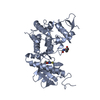 7fhtC 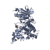 3anrS S: Starting model for refinement C: citing same article ( |
|---|---|
| Similar structure data |
-Links
 PDBj
PDBj
- Assembly
| Deposited unit | 
| ||||||||
|---|---|---|---|---|---|---|---|---|---|
| 1 | 
| ||||||||
| 2 | 
| ||||||||
| 3 | 
| ||||||||
| 4 | 
| ||||||||
| Unit cell |
|
-Components
| #1: Protein | DYRK1A / Dual specificity YAK1-related kinase / HP86 / Protein kinase minibrain homolog / MNBH / hMNB Mass: 42007.391 Da / Num. of mol.: 4 Source method: isolated from a genetically manipulated source Source: (gene. exp.) Homo sapiens (human) / Gene: DYRK1A, DYRK, MNB, MNBH / Production host:  Escherichia coli (E. coli) / References: UniProt: Q13627, dual-specificity kinase Escherichia coli (E. coli) / References: UniProt: Q13627, dual-specificity kinase#2: Chemical | ChemComp-4VZ / (   Mass: 281.351 Da / Num. of mol.: 4 / Source method: obtained synthetically / Formula: C12H11NO3S2 / Feature type: SUBJECT OF INVESTIGATION Mass: 281.351 Da / Num. of mol.: 4 / Source method: obtained synthetically / Formula: C12H11NO3S2 / Feature type: SUBJECT OF INVESTIGATION#3: Chemical | ChemComp-GOL / Glycerol  Mass: 92.094 Da / Num. of mol.: 5 / Source method: obtained synthetically / Formula: C3H8O3 Mass: 92.094 Da / Num. of mol.: 5 / Source method: obtained synthetically / Formula: C3H8O3#4: Water | ChemComp-HOH / | Water Mass: 18.015 Da / Num. of mol.: 252 / Source method: isolated from a natural source / Formula: H2O Mass: 18.015 Da / Num. of mol.: 252 / Source method: isolated from a natural source / Formula: H2OHas ligand of interest | Y | |
|---|
-Experimental details
-Experiment
| Experiment | Method: X-RAY DIFFRACTION / Number of used crystals: 1 |
|---|
- Sample preparation
| Crystal | Density Matthews: 2.62 Å3/Da / Density % sol: 53.09 % |
|---|---|
| Crystal grow | Temperature: 293 K / Method: vapor diffusion, sitting drop Details: 100 mM Bis-Tris propane (pH 8.0), 200 mM Sodium fluoride and 18 % (w/v) PEG 3350 |
-Data collection
| Diffraction | Mean temperature: 100 K / Serial crystal experiment: N |
|---|---|
| Diffraction source | Source: SYNCHROTRON / Site: Photon Factory / Beamline: BL-17A / Wavelength: 0.98 Å |
| Detector | Type: DECTRIS PILATUS3 6M / Detector: PIXEL / Date: Apr 25, 2017 |
| Radiation | Protocol: SINGLE WAVELENGTH / Monochromatic (M) / Laue (L): M / Scattering type: x-ray |
| Radiation wavelength | Wavelength: 0.98 Å / Relative weight: 1 |
| Reflection | Resolution: 2.42→48.12 Å / Num. obs: 67838 / % possible obs: 99.5 % / Redundancy: 6.7 % / Rsym value: 0.15 / Net I/σ(I): 10.2 |
| Reflection shell | Resolution: 2.42→2.48 Å / Num. unique obs: 4466 / Rsym value: 1.432 |
- Processing
| Software |
| ||||||||||||||||||||||||||||||||||||||||||||||||||||||||||||||||||||||||||||||||||||||||||||||||||||||||||||||||||||||||||||||||||||||||||||||||||||||||||||||||||||||||||||||||||||||
|---|---|---|---|---|---|---|---|---|---|---|---|---|---|---|---|---|---|---|---|---|---|---|---|---|---|---|---|---|---|---|---|---|---|---|---|---|---|---|---|---|---|---|---|---|---|---|---|---|---|---|---|---|---|---|---|---|---|---|---|---|---|---|---|---|---|---|---|---|---|---|---|---|---|---|---|---|---|---|---|---|---|---|---|---|---|---|---|---|---|---|---|---|---|---|---|---|---|---|---|---|---|---|---|---|---|---|---|---|---|---|---|---|---|---|---|---|---|---|---|---|---|---|---|---|---|---|---|---|---|---|---|---|---|---|---|---|---|---|---|---|---|---|---|---|---|---|---|---|---|---|---|---|---|---|---|---|---|---|---|---|---|---|---|---|---|---|---|---|---|---|---|---|---|---|---|---|---|---|---|---|---|---|---|
| Refinement | Method to determine structure: MOLECULAR REPLACEMENT Starting model: 3ANR Resolution: 2.42→45 Å / Cor.coef. Fo:Fc: 0.938 / Cor.coef. Fo:Fc free: 0.912 / SU B: 12.91 / SU ML: 0.274 / Cross valid method: THROUGHOUT / ESU R: 0.455 / ESU R Free: 0.287 / Stereochemistry target values: MAXIMUM LIKELIHOOD / Details: HYDROGENS HAVE BEEN USED IF PRESENT IN THE INPUT
| ||||||||||||||||||||||||||||||||||||||||||||||||||||||||||||||||||||||||||||||||||||||||||||||||||||||||||||||||||||||||||||||||||||||||||||||||||||||||||||||||||||||||||||||||||||||
| Solvent computation | Ion probe radii: 0.8 Å / Shrinkage radii: 0.8 Å / VDW probe radii: 1.2 Å / Solvent model: MASK | ||||||||||||||||||||||||||||||||||||||||||||||||||||||||||||||||||||||||||||||||||||||||||||||||||||||||||||||||||||||||||||||||||||||||||||||||||||||||||||||||||||||||||||||||||||||
| Displacement parameters | Biso mean: 52.568 Å2
| ||||||||||||||||||||||||||||||||||||||||||||||||||||||||||||||||||||||||||||||||||||||||||||||||||||||||||||||||||||||||||||||||||||||||||||||||||||||||||||||||||||||||||||||||||||||
| Refinement step | Cycle: 1 / Resolution: 2.42→45 Å
| ||||||||||||||||||||||||||||||||||||||||||||||||||||||||||||||||||||||||||||||||||||||||||||||||||||||||||||||||||||||||||||||||||||||||||||||||||||||||||||||||||||||||||||||||||||||
| Refine LS restraints |
|