 Movie
Movie Controller


+ Open data

- Basic information
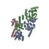
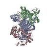
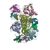
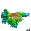
| Entry | Database: PDB / ID: 7fas | ||||||
|---|---|---|---|---|---|---|---|
| Title | VAR2CSA 3D7 ectodomain core region | ||||||
 Components Components | Erythrocyte membrane protein 1, PfEMP1 Red blood cell Red blood cell | ||||||
 Keywords Keywords | CELL ADHESION / Plasmodium falciparum / Complex / CSA binding | ||||||
| Function / homology |  Function and homology information Function and homology informationmodulation by symbiont of host erythrocyte aggregation / infected host cell surface knob / antigenic variation / adhesion of symbiont to microvasculature / cell adhesion molecule binding / cell-cell adhesion / host cell surface receptor binding / host cell plasma membrane / membraneSimilarity search - Function | ||||||
| Biological species |  Plasmodium falciparum (malaria parasite P. falciparum) Plasmodium falciparum (malaria parasite P. falciparum) | ||||||
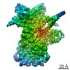
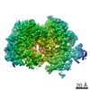
| Method | ELECTRON MICROSCOPY / single particle reconstruction / cryo EM / Resolution: 3.6 Å | ||||||
 Authors Authors | Wang, L. / Zhaoning, W. | ||||||
| Funding support |  China, 1items China, 1items
| ||||||
 Citation Citation | Journal: Cell Discov / Year: 2021 Title: The molecular mechanism of cytoadherence to placental or tumor cells through VAR2CSA from Plasmodium falciparum. Authors: Weiwei Wang / Zhaoning Wang / Xiuna Yang / Yan Gao / Xiang Zhang / Long Cao / Aguang Dai / Jin Sun / Lei Sun / Lubin Jiang / Zhenguo Chen / Lanfeng Wang / | ||||||
| History |
|
- Structure visualization
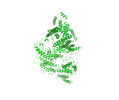
| Movie |
Movie viewer |
|---|---|
| Structure viewer | Molecule: MolmilJmol/JSmol |
- Downloads & links
-Download
| PDBx/mmCIF format | 7fas.cif.gz | 241.5 KB | Display | PDBx/mmCIF format |
|---|---|---|---|---|
| PDB format | pdb7fas.ent.gz | 191.6 KB | Display | PDB format |
| PDBx/mmJSON format | 7fas.json.gz | Tree view | PDBx/mmJSON format | |
| Others |  Other downloads Other downloads |
-Validation report
| Arichive directory | https://data.pdbj.org/pub/pdb/validation_reports/fa/7fasftp://data.pdbj.org/pub/pdb/validation_reports/fa/7fas | HTTPS FTP |
|---|
-Related structure data
| Related structure data |  31505MC 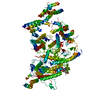 7fapC M: map data used to model this data C: citing same article ( |
|---|---|
| Similar structure data |


-Links
 PDBj
PDBj- Assembly
| Deposited unit | 
|
|---|---|
| 1 |
|
-Components
| #1: Protein | Red blood cell Mass: 228991.188 Da / Num. of mol.: 1 Source method: isolated from a genetically manipulated source Source: (gene. exp.) Plasmodium falciparum (isolate 3D7) (eukaryote)Strain: isolate 3D7 / Gene: PF3D7_1200600 / Production host:   Spodoptera frugiperda (fall armyworm) / References: UniProt: Q8I639 Spodoptera frugiperda (fall armyworm) / References: UniProt: Q8I639 |
|---|---|
| Has ligand of interest | N |
-Experimental details
-Experiment
| Experiment | Method: ELECTRON MICROSCOPY |
|---|---|
| EM experiment | Aggregation state: PARTICLE / 3D reconstruction method: single particle reconstruction |
- Sample preparation
| Component | Name: VAR2CSAPlasmodium falciparum erythrocyte membrane protein 1 Type: COMPLEX / Entity ID: all / Source: RECOMBINANT |
|---|---|
| Molecular weight | Value: 306 kDa/nm / Experimental value: NO |
| Source (natural) | Organism: Plasmodium falciparum 3D7 (eukaryote) |
| Source (recombinant) | Organism: Spodoptera frugiperda (fall armyworm) |
| Buffer solution | pH: 6.5 |
| Specimen | Embedding applied: NO / Shadowing applied: NO / Staining applied: NO / Vitrification applied: YES |
| Vitrification | Cryogen name: ETHANE |
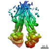
- Electron microscopy imaging
| Experimental equipment |  Model: Titan Krios / Image courtesy: FEI Company |
|---|---|
| Microscopy | Model: FEI TITAN KRIOS |
| Electron gun | Electron source: FIELD EMISSION GUN / Accelerating voltage: 300 kV / Illumination mode: OTHER |
| Electron lens | Mode: BRIGHT FIELDBright-field microscopy |
| Image recording | Electron dose: 52 e/Å2 / Film or detector model: GATAN K2 SUMMIT (4k x 4k) |
- Processing
| Software | Name: PHENIX / Version: 1.17.1_3660: / Classification: refinement | ||||||||||||||||||||||||
|---|---|---|---|---|---|---|---|---|---|---|---|---|---|---|---|---|---|---|---|---|---|---|---|---|---|
| CTF correction | Type: PHASE FLIPPING AND AMPLITUDE CORRECTION | ||||||||||||||||||||||||
| 3D reconstruction | Resolution: 3.6 Å / Resolution method: FSC 0.143 CUT-OFF / Num. of particles: 304160 / Symmetry type: POINT | ||||||||||||||||||||||||
| Atomic model building | Protocol: AB INITIO MODEL | ||||||||||||||||||||||||
| Refine LS restraints |
|
