 Movie
Movie Controller

[English] 日本語
 Yorodumi
- PDB-7f7c: Crystal structure of Non-specific class-C acid phosphatase from S... -
+ Open data

- Basic information
| Entry | Database: PDB / ID: 7f7c | ||||||
|---|---|---|---|---|---|---|---|
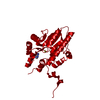
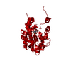
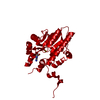
| Title | Crystal structure of Non-specific class-C acid phosphatase from Sphingobium sp. RSMS bound to Adenosine at pH 5.5 | ||||||
 Components Components | Acid phosphatase | ||||||
 Keywords Keywords | HYDROLASE / Non specific Phosphatase / Class c / HAD superfamily. | ||||||
| Function / homology | 5-nucleotidase lipoprotein e(P4) / Acid phosphatase, class B-like / HAD superfamily, subfamily IIIB (Acid phosphatase) / HAD superfamily / cell outer membrane / HAD-like superfamily / ADENOSINE / PHOSPHATE ION / Acid phosphatase Function and homology information Function and homology information | ||||||
| Biological species |  Sphingobium sp. 20006FA (bacteria) Sphingobium sp. 20006FA (bacteria) | ||||||
| Method | X-RAY DIFFRACTION / SYNCHROTRON / AB INITIO PHASING / Resolution: 2.2 Å | ||||||
 Authors Authors | Gaur, N.K. / Kumar, A. / Sunder, S. / Mukhopadhyaya, R. / Makde, R.D. | ||||||
 Citation Citation | Journal: To Be Published Title: Non-Specific Class-c acidphosphatase from Sphingobium sp. RSMS Authors: Gaur, N.K. / Kumar, A. / Sunder, S. / Mukhopadhyaya, R. / Makde, R.D. | ||||||
| History |
|
- Structure visualization
| Structure viewer | Molecule: MolmilJmol/JSmol |
|---|
- Downloads & links
-Download
| PDBx/mmCIF format | 7f7c.cif.gz | 178.5 KB | Display | PDBx/mmCIF format |
|---|---|---|---|---|
| PDB format | pdb7f7c.ent.gz | 121.7 KB | Display | PDB format |
| PDBx/mmJSON format | 7f7c.json.gz | Tree view | PDBx/mmJSON format | |
| Others |  Other downloads Other downloads |
-Validation report
| Arichive directory | https://data.pdbj.org/pub/pdb/validation_reports/f7/7f7cftp://data.pdbj.org/pub/pdb/validation_reports/f7/7f7c | HTTPS FTP |
|---|
-Related structure data

-Links
 PDBj
PDBj
- Assembly
| Deposited unit | 
| ||||||||||
|---|---|---|---|---|---|---|---|---|---|---|---|
| 1 | 
| ||||||||||
| Unit cell |
| ||||||||||
| Components on special symmetry positions |
|
-Components
| #1: Protein | Mass: 31206.871 Da / Num. of mol.: 1 Source method: isolated from a genetically manipulated source Source: (gene. exp.) Sphingobium sp. 20006FA (bacteria) / Gene: A8O16_10785 / Production host: Escherichia coli (E. coli) / Strain (production host): BL 21 DE3 / References: UniProt: A0A197BYF0 |
|---|---|
| #2: Chemical | ChemComp-ADN / Adenosine  Mass: 267.241 Da / Num. of mol.: 1 / Source method: obtained synthetically / Formula: C10H13N5O4 / Feature type: SUBJECT OF INVESTIGATION Mass: 267.241 Da / Num. of mol.: 1 / Source method: obtained synthetically / Formula: C10H13N5O4 / Feature type: SUBJECT OF INVESTIGATION |
| #3: Chemical | ChemComp-PO4 / Phosphate  Mass: 94.971 Da / Num. of mol.: 1 / Source method: obtained synthetically / Formula: PO4 / Feature type: SUBJECT OF INVESTIGATION Mass: 94.971 Da / Num. of mol.: 1 / Source method: obtained synthetically / Formula: PO4 / Feature type: SUBJECT OF INVESTIGATION |
| #4: Chemical | ChemComp-MG /   Mass: 24.305 Da / Num. of mol.: 1 / Source method: isolated from a natural source / Formula: Mg Mass: 24.305 Da / Num. of mol.: 1 / Source method: isolated from a natural source / Formula: Mg |
| #5: Water | ChemComp-HOH / Water Mass: 18.015 Da / Num. of mol.: 127 / Source method: isolated from a natural source / Formula: H2O Mass: 18.015 Da / Num. of mol.: 127 / Source method: isolated from a natural source / Formula: H2O |
| Has ligand of interest | Y |
-Experimental details
-Experiment
| Experiment | Method: X-RAY DIFFRACTION / Number of used crystals: 1 |
|---|
- Sample preparation
| Crystal | Density Matthews: 3.17 Å3/Da / Density % sol: 66.66 % / Description: Hexagonal Crystals |
|---|---|
| Crystal grow | Temperature: 294 K / Method: microbatch Details: 1M Ammonium sulphate, 0.1 M Bis-tris pH 5.5, 1% PEG 3350 |
-Data collection
| Diffraction | Mean temperature: 100 K / Serial crystal experiment: N |
|---|---|
| Diffraction source | Source: SYNCHROTRON / Site: RRCAT INDUS-2  / Beamline: PX-BL21 / Wavelength: 0.9795 Å / Beamline: PX-BL21 / Wavelength: 0.9795 Å |
| Detector | Type: MAR scanner 300 mm plate / Detector: IMAGE PLATE / Date: Oct 6, 2018 |
| Radiation | Protocol: SINGLE WAVELENGTH / Monochromatic (M) / Laue (L): M / Scattering type: x-ray |
| Radiation wavelength | Wavelength: 0.9795 Å / Relative weight: 1 |
| Reflection | Resolution: 2.2→48.5 Å / Num. obs: 20831 / % possible obs: 100 % / Redundancy: 13.8 % / Biso Wilson estimate: 30.79 Å2 / CC1/2: 0.997 / Net I/σ(I): 10.5 |
| Reflection shell | Resolution: 2.2→2.27 Å / Num. unique obs: 1758 / CC1/2: 0.686 |
- Processing
| Software |
| |||||||||||||||||||||||||||||||||||||||||||||||||||||||||||||||
|---|---|---|---|---|---|---|---|---|---|---|---|---|---|---|---|---|---|---|---|---|---|---|---|---|---|---|---|---|---|---|---|---|---|---|---|---|---|---|---|---|---|---|---|---|---|---|---|---|---|---|---|---|---|---|---|---|---|---|---|---|---|---|---|---|
| Refinement | Method to determine structure: AB INITIO PHASING / Resolution: 2.2→48.5 Å / SU ML: 0.2296 / Cross valid method: FREE R-VALUE / σ(F): 1.34 / Phase error: 22.767 Stereochemistry target values: GeoStd + Monomer Library + CDL v1.2
| |||||||||||||||||||||||||||||||||||||||||||||||||||||||||||||||
| Solvent computation | Shrinkage radii: 0.9 Å / VDW probe radii: 1.11 Å / Solvent model: FLAT BULK SOLVENT MODEL | |||||||||||||||||||||||||||||||||||||||||||||||||||||||||||||||
| Displacement parameters | Biso mean: 36.44 Å2 | |||||||||||||||||||||||||||||||||||||||||||||||||||||||||||||||
| Refinement step | Cycle: LAST / Resolution: 2.2→48.5 Å
| |||||||||||||||||||||||||||||||||||||||||||||||||||||||||||||||
| Refine LS restraints |
| |||||||||||||||||||||||||||||||||||||||||||||||||||||||||||||||
| LS refinement shell |
| |||||||||||||||||||||||||||||||||||||||||||||||||||||||||||||||
| Refinement TLS params. | Method: refined / Origin x: 4.35166904967 Å / Origin y: -20.8422872627 Å / Origin z: 29.0058386633 Å
| |||||||||||||||||||||||||||||||||||||||||||||||||||||||||||||||
| Refinement TLS group | Selection details: all |
