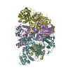
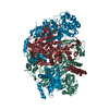
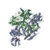
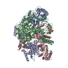
- PDB-7f4y: Crystal structure of replisomal dimer of DNA polymerase from bact... -
+
Open data
ID or keywords:
Loading...
-
Basic information
Entry
Database: PDB / ID: 7f4y
Title
Crystal structure of replisomal dimer of DNA polymerase from bacteriophage RB69 with DNA duplexes
Components
DNA (5'-D(*GP*AP*GP*CP*GP*GP*AP*CP*TP*GP*CP*TP*TP*AP*C)-3')
DNA (5'-D(*TP*CP*AP*AP*GP*TP*AP*AP*GP*CP*AP*GP*TP*CP*CP*GP*CP*TP*C)-3')
DNA polymerase
Keywords
TRANSFERASE/DNA / TRANSFERASE-DNA complex
Function / homology
Function and homology information
bidirectional double-stranded viral DNA replication / nucleotide-excision repair, DNA gap filling / DNA replication proofreading / 3'-5'-DNA exonuclease activity / Hydrolases; Acting on ester bonds; Exodeoxyribonucleases producing 5'-phosphomonoesters / SOS response / 3'-5' exonuclease activity / base-excision repair, gap-filling / DNA-directed DNA polymerase / DNA-directed DNA polymerase activity ...bidirectional double-stranded viral DNA replication / nucleotide-excision repair, DNA gap filling / DNA replication proofreading / 3'-5'-DNA exonuclease activity / Hydrolases; Acting on ester bonds; Exodeoxyribonucleases producing 5'-phosphomonoesters / SOS response / 3'-5' exonuclease activity / base-excision repair, gap-filling / DNA-directed DNA polymerase / DNA-directed DNA polymerase activity / nucleotide binding / DNA binding / metal ion binding Similarity search - Function
DNA-directed DNA polymerase T4 type / DNA polymerase family B signature. / DNA-directed DNA polymerase, family B, conserved site / DNA polymerase family B / DNA polymerase family B, exonuclease domain / DNA-directed DNA polymerase, family B, exonuclease domain / DNA-directed DNA polymerase, family B, multifunctional domain / DNA polymerase, palm domain superfamily / DNA polymerase type-B family / DNA-directed DNA polymerase, family B ...DNA-directed DNA polymerase T4 type / DNA polymerase family B signature. / DNA-directed DNA polymerase, family B, conserved site / DNA polymerase family B / DNA polymerase family B, exonuclease domain / DNA-directed DNA polymerase, family B, exonuclease domain / DNA-directed DNA polymerase, family B, multifunctional domain / DNA polymerase, palm domain superfamily / DNA polymerase type-B family / DNA-directed DNA polymerase, family B / Ribonuclease H superfamily / Ribonuclease H-like superfamily / DNA/RNA polymerase superfamily Similarity search - Domain/homology
GUANOSINE-5'-MONOPHOSPHATE / 2'-DEOXYURIDINE 5'-ALPHA,BETA-IMIDO-TRIPHOSPHATE / DNA / DNA (> 10) / DNA-directed DNA polymerase Similarity search - Component
A: DNA polymerase T: DNA (5'-D(*TP*CP*AP*AP*GP*TP*AP*AP*GP*CP*AP*GP*TP*CP*CP*GP*CP*TP*C)-3') P: DNA (5'-D(*GP*AP*GP*CP*GP*GP*AP*CP*TP*GP*CP*TP*TP*AP*C)-3') B: DNA polymerase S: DNA (5'-D(*TP*CP*AP*AP*GP*TP*AP*AP*GP*CP*AP*GP*TP*CP*CP*GP*CP*TP*C)-3') Q: DNA (5'-D(*GP*AP*GP*CP*GP*GP*AP*CP*TP*GP*CP*TP*TP*AP*C)-3') hetero molecules
In the structure databanks used in Yorodumi, some data are registered as the other names, "COVID-19 virus" and "2019-nCoV". Here are the details of the virus and the list of structure data.
Jan 31, 2019. EMDB accession codes are about to change! (news from PDBe EMDB page)
EMDB accession codes are about to change! (news from PDBe EMDB page)
The allocation of 4 digits for EMDB accession codes will soon come to an end. Whilst these codes will remain in use, new EMDB accession codes will include an additional digit and will expand incrementally as the available range of codes is exhausted. The current 4-digit format prefixed with “EMD-” (i.e. EMD-XXXX) will advance to a 5-digit format (i.e. EMD-XXXXX), and so on. It is currently estimated that the 4-digit codes will be depleted around Spring 2019, at which point the 5-digit format will come into force.
The EM Navigator/Yorodumi systems omit the EMD- prefix.
Related info.:Q: What is EMD? / ID/Accession-code notation in Yorodumi/EM Navigator
Yorodumi is a browser for structure data from EMDB, PDB, SASBDB, etc.
This page is also the successor to EM Navigator detail page, and also detail information page/front-end page for Omokage search.
The word "yorodu" (or yorozu) is an old Japanese word meaning "ten thousand". "mi" (miru) is to see.
Related info.:EMDB / PDB / SASBDB / Comparison of 3 databanks / Yorodumi Search / Aug 31, 2016. New EM Navigator & Yorodumi / Yorodumi Papers / Jmol/JSmol / Function and homology information / Changes in new EM Navigator and Yorodumi
 Movie
Movie Controller

 Yorodumi
 Open data
 Basic information
 Components
 Keywords
 Function and homology information
 Hydrolases; Acting on ester bonds; Exodeoxyribonucleases producing 5'-phosphomonoesters /
  Enterobacteria phage RB69 (virus)
 Authors
 Korea, Republic Of, 1items
  Citation
 Structure visualization
 Downloads & links
 Other downloads


 PDBj
PDBj


 Assembly
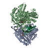

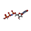


 Mass: 467.157 Da / Num. of mol.: 1 / Source method: obtained synthetically / Formula: C9H16N3O13P3
 Mass: 40.078 Da / Num. of mol.: 2 / Source method: obtained synthetically / Formula: Ca
 Mass: 363.221 Da / Num. of mol.: 2 / Source method: obtained synthetically / Formula: C10H14N5O8P
 Mass: 24.305 Da / Num. of mol.: 1 / Source method: obtained synthetically / Formula: Mg
 Sample preparation
 / Beamline: AR-NW12A / Wavelength: 1 Å
 Processing