 Movie
Movie Controller


+ Open data

- Basic information
| Entry | Database: PDB / ID: 7ezn | ||||||
|---|---|---|---|---|---|---|---|
| Title | Crystal structure of CnYvh1 complex with vanadate | ||||||
 Components Components | Protein-tyrosine-phosphatase Protein tyrosine phosphatase Protein tyrosine phosphatase | ||||||
 Keywords Keywords | HYDROLASE / DUSP / phosphatase / inhibitor | ||||||
| Function / homology |  Function and homology information Function and homology informationprotein tyrosine/serine/threonine phosphatase activity / protein tyrosine phosphatase activitySimilarity search - Function | ||||||
| Biological species |  Cryptococcus neoformans var. grubii serotype A (fungus) Cryptococcus neoformans var. grubii serotype A (fungus) | ||||||
| Method | X-RAY DIFFRACTION / SYNCHROTRON / MOLECULAR REPLACEMENT / Resolution: 1.8 Å | ||||||
 Authors Authors | Choi, M.K. / Yoo, Y. | ||||||
 Citation Citation | Journal: To Be Published Title: Crystal structure of CnYvh1 complex with vanadate Authors: Choi, M.K. / Yoo, Y. #1: Journal: Acta Crystallogr D Biol Crystallogr / Year: 2014Title: The family-wide structure and function of human dual-specificity protein phosphatases. Authors: Jeong, D.G. / Wei, C.H. / Ku, B. / Jeon, T.J. / Chien, P.N. / Kim, J.K. / Park, S.Y. / Hwang, H.S. / Ryu, S.Y. / Park, H. / Kim, D.S. / Kim, S.J. / Ryu, S.E. | ||||||
| History |
|
- Structure visualization
| Structure viewer | Molecule: MolmilJmol/JSmol |
|---|
- Downloads & links
-Download
| PDBx/mmCIF format | 7ezn.cif.gz | 52.6 KB | Display | PDBx/mmCIF format |
|---|---|---|---|---|
| PDB format | pdb7ezn.ent.gz | 34.1 KB | Display | PDB format |
| PDBx/mmJSON format | 7ezn.json.gz | Tree view | PDBx/mmJSON format | |
| Others |  Other downloads Other downloads |
-Validation report
| Arichive directory | https://data.pdbj.org/pub/pdb/validation_reports/ez/7eznftp://data.pdbj.org/pub/pdb/validation_reports/ez/7ezn | HTTPS FTP |
|---|
-Related structure data
| Related structure data | 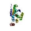 4ki9S S: Starting model for refinement |
|---|---|
| Similar structure data |
-Links
 PDBj
PDBj


- Assembly
| Deposited unit | 
| ||||||||||||
|---|---|---|---|---|---|---|---|---|---|---|---|---|---|
| 1 |
| ||||||||||||
| Unit cell |
| ||||||||||||
| Components on special symmetry positions |
|
-Components
| #1: Protein | Protein tyrosine phosphatase Mass: 22754.635 Da / Num. of mol.: 1 Source method: isolated from a genetically manipulated source Source: (gene. exp.) Cryptococcus neoformans var. grubii serotype A (strain H99 / ATCC 208821 / CBS 10515 / FGSC 9487) (fungus)Strain: H99 / ATCC 208821 / CBS 10515 / FGSC 9487 / Gene: CNAG_01203 / Production host:  Escherichia coli BL21(DE3) (bacteria) / References: UniProt: J9VTB5, protein-tyrosine-phosphatase Escherichia coli BL21(DE3) (bacteria) / References: UniProt: J9VTB5, protein-tyrosine-phosphatase |
|---|---|
| #2: Chemical | ChemComp-VO4 / Vanadate  Mass: 114.939 Da / Num. of mol.: 1 / Source method: obtained synthetically / Formula: VO4 / Feature type: SUBJECT OF INVESTIGATION Mass: 114.939 Da / Num. of mol.: 1 / Source method: obtained synthetically / Formula: VO4 / Feature type: SUBJECT OF INVESTIGATION |
| #3: Chemical | ChemComp-EDO / Ethylene glycol  Mass: 62.068 Da / Num. of mol.: 1 / Source method: obtained synthetically / Formula: C2H6O2 Mass: 62.068 Da / Num. of mol.: 1 / Source method: obtained synthetically / Formula: C2H6O2 |
| #4: Water | ChemComp-HOH / Water Mass: 18.015 Da / Num. of mol.: 76 / Source method: isolated from a natural source / Formula: H2O Mass: 18.015 Da / Num. of mol.: 76 / Source method: isolated from a natural source / Formula: H2O |
| Has ligand of interest | Y |
-Experimental details
-Experiment
| Experiment | Method: X-RAY DIFFRACTION / Number of used crystals: 1 |
|---|
- Sample preparation
| Crystal | Density Matthews: 2.9 Å3/Da / Density % sol: 57.53 % |
|---|---|
| Crystal grow | Temperature: 298 K / Method: vapor diffusion, sitting drop / pH: 8.5 Details: 0.16 M Magnesium chloride, 0.08 M Tris-HCl pH 8.5, 24 % (w/v) PEG 4000, 20 % glycerol |
-Data collection
| Diffraction | Mean temperature: 100 K / Serial crystal experiment: N |
|---|---|
| Diffraction source | Source: SYNCHROTRON / Site: PAL/PLS  / Beamline: 7A (6B, 6C1) / Wavelength: 0.97 Å / Beamline: 7A (6B, 6C1) / Wavelength: 0.97 Å |
| Detector | Type: ADSC QUANTUM 270 / Detector: CCD / Date: Jul 26, 2018 |
| Radiation | Protocol: SINGLE WAVELENGTH / Monochromatic (M) / Laue (L): M / Scattering type: x-ray |
| Radiation wavelength | Wavelength: 0.97 Å / Relative weight: 1 |
| Reflection | Resolution: 1.8→33.1 Å / Num. obs: 24183 / % possible obs: 94.58 % / Redundancy: 2 % / Biso Wilson estimate: 29.88 Å2 / CC1/2: 1 / Rmerge(I) obs: 0.01125 / Net I/σ(I): 33.71 |
| Reflection shell | Resolution: 1.8→1.864 Å / Rmerge(I) obs: 0.2128 / Num. unique obs: 1709 / CC1/2: 0.926 |
- Processing
| Software |
| ||||||||||||||||||||||||||||||||||||||||||||||||||||||||||||||||||||||||||||||||||||||||||||||||||
|---|---|---|---|---|---|---|---|---|---|---|---|---|---|---|---|---|---|---|---|---|---|---|---|---|---|---|---|---|---|---|---|---|---|---|---|---|---|---|---|---|---|---|---|---|---|---|---|---|---|---|---|---|---|---|---|---|---|---|---|---|---|---|---|---|---|---|---|---|---|---|---|---|---|---|---|---|---|---|---|---|---|---|---|---|---|---|---|---|---|---|---|---|---|---|---|---|---|---|---|
| Refinement | Method to determine structure: MOLECULAR REPLACEMENT Starting model: 4ki9 Resolution: 1.8→33.1 Å / SU ML: 0.1345 / Cross valid method: FREE R-VALUE / σ(F): 1.35 / Phase error: 22.6013 Stereochemistry target values: GeoStd + Monomer Library + CDL v1.2
| ||||||||||||||||||||||||||||||||||||||||||||||||||||||||||||||||||||||||||||||||||||||||||||||||||
| Solvent computation | Shrinkage radii: 0.9 Å / VDW probe radii: 1.11 Å / Solvent model: FLAT BULK SOLVENT MODEL | ||||||||||||||||||||||||||||||||||||||||||||||||||||||||||||||||||||||||||||||||||||||||||||||||||
| Displacement parameters | Biso mean: 32.94 Å2 | ||||||||||||||||||||||||||||||||||||||||||||||||||||||||||||||||||||||||||||||||||||||||||||||||||
| Refinement step | Cycle: LAST / Resolution: 1.8→33.1 Å
| ||||||||||||||||||||||||||||||||||||||||||||||||||||||||||||||||||||||||||||||||||||||||||||||||||
| Refine LS restraints |
| ||||||||||||||||||||||||||||||||||||||||||||||||||||||||||||||||||||||||||||||||||||||||||||||||||
| LS refinement shell |
|
