 Movie
Movie Controller

[English] 日本語
 Yorodumi
- PDB-7ex0: Crystal structure of Ebinur Lake virus cap snatching endonuclease... -
+ Open data

- Basic information
| Entry | Database: PDB / ID: 7ex0 | |||||||||
|---|---|---|---|---|---|---|---|---|---|---|
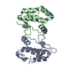
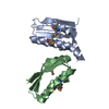
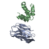
| Title | Crystal structure of Ebinur Lake virus cap snatching endonuclease (K108A mutant) | |||||||||
 Components Components | Replicase RNA-dependent RNA polymerase RNA-dependent RNA polymerase | |||||||||
 Keywords Keywords | HYDROLASE / endonuclease | |||||||||
| Function / homology |  Function and homology information Function and homology informationhost cell endoplasmic reticulum / host cell endoplasmic reticulum-Golgi intermediate compartment / host cell Golgi apparatus / RNA-directed RNA polymerase / viral RNA genome replication / RNA-dependent RNA polymerase activity / DNA-templated transcription / metal ion bindingSimilarity search - Function | |||||||||
| Biological species |  Abbey lake orthobunyavirus Abbey lake orthobunyavirus | |||||||||
| Method | X-RAY DIFFRACTION / SYNCHROTRON / MOLECULAR REPLACEMENT / Resolution: 2.07 Å | |||||||||
 Authors Authors | Kuang, W. / Hu, Z. / Gong, P. | |||||||||
| Funding support |  China, 2items China, 2items
| |||||||||
 Citation Citation | Journal: J.Virol. / Year: 2022 Title: Insights into Two-Metal-Ion Catalytic Mechanism of Cap-Snatching Endonuclease of Ebinur Lake Virus in Bunyavirales. Authors: Kuang, W. / Zhang, H. / Cai, Y. / Zhang, G. / Deng, F. / Li, H. / Hu, Z. / Guo, Y. / Wang, M. / Zhou, Y. / Gong, P. | |||||||||
| History |
|
- Structure visualization
| Structure viewer | Molecule: MolmilJmol/JSmol |
|---|
- Downloads & links
-Download
| PDBx/mmCIF format | 7ex0.cif.gz | 92.1 KB | Display | PDBx/mmCIF format |
|---|---|---|---|---|
| PDB format | pdb7ex0.ent.gz | 66.5 KB | Display | PDB format |
| PDBx/mmJSON format | 7ex0.json.gz | Tree view | PDBx/mmJSON format | |
| Others |  Other downloads Other downloads |
-Validation report
| Arichive directory | https://data.pdbj.org/pub/pdb/validation_reports/ex/7ex0ftp://data.pdbj.org/pub/pdb/validation_reports/ex/7ex0 | HTTPS FTP |
|---|
-Related structure data
| Related structure data |  7ewuC  7ewvC  7ewwC  7ewxC  7ewyC  7ewzC  7ex1C  2xi5S S: Starting model for refinement C: citing same article ( |
|---|---|
| Similar structure data |

-Links
 PDBj
PDBj- Assembly
| Deposited unit | 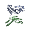
| ||||||||
|---|---|---|---|---|---|---|---|---|---|
| 1 | 
| ||||||||
| 2 | 
| ||||||||
| Unit cell |
|
-Components
| #1: Protein | RNA-dependent RNA polymerase / Transcriptase / L protein Mass: 25123.773 Da / Num. of mol.: 2 / Fragment: N-terminal endonuclease domain / Mutation: K108A Source method: isolated from a genetically manipulated source Source: (gene. exp.) Abbey lake orthobunyavirus / Strain: Cu20-XJ / Gene: RdRp / Plasmid: pET28 / Production host:  Escherichia coli BL21(DE3) (bacteria) Escherichia coli BL21(DE3) (bacteria)References: UniProt: A0A059WLS9, RNA-directed RNA polymerase#2: Chemical | ChemComp-MN /   Mass: 54.938 Da / Num. of mol.: 4 / Source method: obtained synthetically / Formula: Mn Mass: 54.938 Da / Num. of mol.: 4 / Source method: obtained synthetically / Formula: Mn#3: Water | ChemComp-HOH / | Water Mass: 18.015 Da / Num. of mol.: 191 / Source method: isolated from a natural source / Formula: H2O Mass: 18.015 Da / Num. of mol.: 191 / Source method: isolated from a natural source / Formula: H2OHas ligand of interest | N | |
|---|
-Experimental details
-Experiment
| Experiment | Method: X-RAY DIFFRACTION / Number of used crystals: 1 |
|---|
- Sample preparation
| Crystal | Density Matthews: 2.39 Å3/Da / Density % sol: 48.5 % / Mosaicity: 0.883 ° |
|---|---|
| Crystal grow | Temperature: 289 K / Method: evaporation / pH: 5.1 / Details: PEG 3350 |
-Data collection
| Diffraction | Mean temperature: 100 K / Serial crystal experiment: N | |||||||||||||||||||||||||||||||||||||||||||||||||||||||||||||||||||||||||||||||||||||||||||||||||||
|---|---|---|---|---|---|---|---|---|---|---|---|---|---|---|---|---|---|---|---|---|---|---|---|---|---|---|---|---|---|---|---|---|---|---|---|---|---|---|---|---|---|---|---|---|---|---|---|---|---|---|---|---|---|---|---|---|---|---|---|---|---|---|---|---|---|---|---|---|---|---|---|---|---|---|---|---|---|---|---|---|---|---|---|---|---|---|---|---|---|---|---|---|---|---|---|---|---|---|---|---|
| Diffraction source | Source: SYNCHROTRON / Site: SSRF / Beamline: BL17U / Wavelength: 0.9792 Å | |||||||||||||||||||||||||||||||||||||||||||||||||||||||||||||||||||||||||||||||||||||||||||||||||||
| Detector | Type: DECTRIS EIGER X 16M / Detector: PIXEL / Date: Dec 8, 2019 | |||||||||||||||||||||||||||||||||||||||||||||||||||||||||||||||||||||||||||||||||||||||||||||||||||
| Radiation | Protocol: SINGLE WAVELENGTH / Monochromatic (M) / Laue (L): M / Scattering type: x-ray | |||||||||||||||||||||||||||||||||||||||||||||||||||||||||||||||||||||||||||||||||||||||||||||||||||
| Radiation wavelength | Wavelength: 0.9792 Å / Relative weight: 1 | |||||||||||||||||||||||||||||||||||||||||||||||||||||||||||||||||||||||||||||||||||||||||||||||||||
| Reflection | Resolution: 2.07→50 Å / Num. obs: 29345 / % possible obs: 99.8 % / Redundancy: 3.6 % / Biso Wilson estimate: 35.91 Å2 / Rmerge(I) obs: 0.04 / Rpim(I) all: 0.024 / Rrim(I) all: 0.047 / Χ2: 0.935 / Net I/σ(I): 15.3 / Num. measured all: 104989 | |||||||||||||||||||||||||||||||||||||||||||||||||||||||||||||||||||||||||||||||||||||||||||||||||||
| Reflection shell | Diffraction-ID: 1
|
- Processing
| Software |
| |||||||||||||||||||||||||||||||||||||||||||||||||||||||||||||||||||||||||||||
|---|---|---|---|---|---|---|---|---|---|---|---|---|---|---|---|---|---|---|---|---|---|---|---|---|---|---|---|---|---|---|---|---|---|---|---|---|---|---|---|---|---|---|---|---|---|---|---|---|---|---|---|---|---|---|---|---|---|---|---|---|---|---|---|---|---|---|---|---|---|---|---|---|---|---|---|---|---|---|
| Refinement | Method to determine structure: MOLECULAR REPLACEMENT Starting model: 2XI5 Resolution: 2.07→30.21 Å / SU ML: 0.28 / Cross valid method: FREE R-VALUE / σ(F): 1.35 / Phase error: 26.42 / Stereochemistry target values: ML
| |||||||||||||||||||||||||||||||||||||||||||||||||||||||||||||||||||||||||||||
| Solvent computation | Shrinkage radii: 0.9 Å / VDW probe radii: 1.11 Å / Solvent model: FLAT BULK SOLVENT MODEL | |||||||||||||||||||||||||||||||||||||||||||||||||||||||||||||||||||||||||||||
| Displacement parameters | Biso max: 124.69 Å2 / Biso mean: 41.8006 Å2 / Biso min: 20.48 Å2 | |||||||||||||||||||||||||||||||||||||||||||||||||||||||||||||||||||||||||||||
| Refinement step | Cycle: final / Resolution: 2.07→30.21 Å
| |||||||||||||||||||||||||||||||||||||||||||||||||||||||||||||||||||||||||||||
| LS refinement shell | Refine-ID: X-RAY DIFFRACTION / Rfactor Rfree error: 0 / Total num. of bins used: 10
|
