 Movie
Movie Controller

[English] 日本語
 Yorodumi
- PDB-7eus: Crystal structures of 2-oxoglutarate dependent dioxygenase (CTB9)... -
+ Open data

- Basic information
| Entry | Database: PDB / ID: 7eus | |||||||||||||||
|---|---|---|---|---|---|---|---|---|---|---|---|---|---|---|---|---|
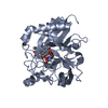
| Title | Crystal structures of 2-oxoglutarate dependent dioxygenase (CTB9) from Cercospora sp. JNU001 | |||||||||||||||
 Components Components | 2-oxoglutarate (2-OG)-dependent dioxygenase | |||||||||||||||
 Keywords Keywords |  OXIDOREDUCTASE / 2-oxoglutarate-dependent dioxygenase / cercosporin OXIDOREDUCTASE / 2-oxoglutarate-dependent dioxygenase / cercosporin | |||||||||||||||
| Function / homology | COPPER (II) ION Function and homology information Function and homology information | |||||||||||||||
| Biological species |  Cercospora sojina (fungus) Cercospora sojina (fungus) | |||||||||||||||
| Method | X-RAY DIFFRACTION / SYNCHROTRON / SAD / Resolution: 2.3 Å | |||||||||||||||
 Authors Authors | Hou, X.D. / Liu, X.Z. / Yuan, Z.B. / Rao, Y.J. | |||||||||||||||
| Funding support |  China, 4items China, 4items
| |||||||||||||||
 Citation Citation | Journal: Acs Catalysis / Year: 2022 Title: Molecular Basis of the Unusual Seven-Membered Methylenedioxy Bridge Formation Catalyzed by Fe(II)/alpha-KG-Dependent Oxygenase CTB9 Authors: Liu, X.Z. / Yuan, Z.B. / Su, H. / Hou, X.D. / Deng, Z.W. / Xu, H.B. / Guo, B.D. / Yin, D. / Sheng, X. / Rao, Y.J. | |||||||||||||||
| History |
|
- Structure visualization
| Structure viewer | Molecule: MolmilJmol/JSmol |
|---|
- Downloads & links
-Download
| PDBx/mmCIF format | 7eus.cif.gz | 133 KB | Display | PDBx/mmCIF format |
|---|---|---|---|---|
| PDB format | pdb7eus.ent.gz | 107.6 KB | Display | PDB format |
| PDBx/mmJSON format | 7eus.json.gz | Tree view | PDBx/mmJSON format | |
| Others |  Other downloads Other downloads |
-Validation report
| Arichive directory | https://data.pdbj.org/pub/pdb/validation_reports/eu/7eusftp://data.pdbj.org/pub/pdb/validation_reports/eu/7eus | HTTPS FTP |
|---|
-Related structure data

-Links
 PDBj
PDBj- Assembly
| Deposited unit | 
| ||||||||
|---|---|---|---|---|---|---|---|---|---|
| 1 | 
| ||||||||
| 2 | 
| ||||||||
| Unit cell |
|
-Components
| #1: Protein | Mass: 39008.090 Da / Num. of mol.: 2 Source method: isolated from a genetically manipulated source Source: (gene. exp.) Cercospora sojina (fungus) / Production host:  Escherichia coli (E. coli) Escherichia coli (E. coli)#2: Chemical | Copper  Mass: 63.546 Da / Num. of mol.: 2 / Source method: obtained synthetically / Formula: Cu / Feature type: SUBJECT OF INVESTIGATION Mass: 63.546 Da / Num. of mol.: 2 / Source method: obtained synthetically / Formula: Cu / Feature type: SUBJECT OF INVESTIGATION#3: Chemical | ChemComp-GOL / | Glycerol  Mass: 92.094 Da / Num. of mol.: 1 / Source method: obtained synthetically / Formula: C3H8O3 / Feature type: SUBJECT OF INVESTIGATION Mass: 92.094 Da / Num. of mol.: 1 / Source method: obtained synthetically / Formula: C3H8O3 / Feature type: SUBJECT OF INVESTIGATION#4: Water | ChemComp-HOH / | Water Mass: 18.015 Da / Num. of mol.: 208 / Source method: isolated from a natural source / Formula: H2O Mass: 18.015 Da / Num. of mol.: 208 / Source method: isolated from a natural source / Formula: H2OHas ligand of interest | Y | |
|---|
-Experimental details
-Experiment
| Experiment | Method: X-RAY DIFFRACTION / Number of used crystals: 1 |
|---|
- Sample preparation
| Crystal | Density Matthews: 2.18 Å3/Da / Density % sol: 43.64 % |
|---|---|
| Crystal grow | Temperature: 291 K / Method: vapor diffusion, sitting drop / pH: 6.5 Details: 0.1 mM Bis-Tris pH 6.5, 3% (v/v) glycerol, 25% (w/v) PEG 3350 |
-Data collection
| Diffraction | Mean temperature: 100 K / Serial crystal experiment: N | |||||||||||||||||||||||||||||||||||||||||||||||||||||||||||||||||||||||||||||||||||||||||||||||||||
|---|---|---|---|---|---|---|---|---|---|---|---|---|---|---|---|---|---|---|---|---|---|---|---|---|---|---|---|---|---|---|---|---|---|---|---|---|---|---|---|---|---|---|---|---|---|---|---|---|---|---|---|---|---|---|---|---|---|---|---|---|---|---|---|---|---|---|---|---|---|---|---|---|---|---|---|---|---|---|---|---|---|---|---|---|---|---|---|---|---|---|---|---|---|---|---|---|---|---|---|---|
| Diffraction source | Source: SYNCHROTRON / Site: NFPSS / Beamline: BL19U1 / Wavelength: 0.979 Å | |||||||||||||||||||||||||||||||||||||||||||||||||||||||||||||||||||||||||||||||||||||||||||||||||||
| Detector | Type: DECTRIS PILATUS3 6M / Detector: PIXEL / Date: Jul 24, 2020 | |||||||||||||||||||||||||||||||||||||||||||||||||||||||||||||||||||||||||||||||||||||||||||||||||||
| Radiation | Protocol: SINGLE WAVELENGTH / Monochromatic (M) / Laue (L): M / Scattering type: x-ray | |||||||||||||||||||||||||||||||||||||||||||||||||||||||||||||||||||||||||||||||||||||||||||||||||||
| Radiation wavelength | Wavelength: 0.979 Å / Relative weight: 1 | |||||||||||||||||||||||||||||||||||||||||||||||||||||||||||||||||||||||||||||||||||||||||||||||||||
| Reflection | Resolution: 2.29→50 Å / Num. obs: 28264 / % possible obs: 98.6 % / Redundancy: 6.6 % / Rmerge(I) obs: 0.125 / Rpim(I) all: 0.051 / Rrim(I) all: 0.135 / Χ2: 0.878 / Net I/σ(I): 6.1 | |||||||||||||||||||||||||||||||||||||||||||||||||||||||||||||||||||||||||||||||||||||||||||||||||||
| Reflection shell | Diffraction-ID: 1
|
- Processing
| Software |
| ||||||||||||||||||||||||||||||||||||||||||||||||||||||||||||
|---|---|---|---|---|---|---|---|---|---|---|---|---|---|---|---|---|---|---|---|---|---|---|---|---|---|---|---|---|---|---|---|---|---|---|---|---|---|---|---|---|---|---|---|---|---|---|---|---|---|---|---|---|---|---|---|---|---|---|---|---|---|
| Refinement | Method to determine structure: SAD / Resolution: 2.3→42.62 Å / Cor.coef. Fo:Fc: 0.947 / Cor.coef. Fo:Fc free: 0.924 / SU B: 5.798 / SU ML: 0.143 / Cross valid method: THROUGHOUT / σ(F): 0 / ESU R: 0.407 / ESU R Free: 0.233 / Stereochemistry target values: MAXIMUM LIKELIHOOD Details: HYDROGENS HAVE BEEN ADDED IN THE RIDING POSITIONS U VALUES : REFINED INDIVIDUALLY
| ||||||||||||||||||||||||||||||||||||||||||||||||||||||||||||
| Solvent computation | Ion probe radii: 0.8 Å / Shrinkage radii: 0.8 Å / VDW probe radii: 1.2 Å / Solvent model: MASK | ||||||||||||||||||||||||||||||||||||||||||||||||||||||||||||
| Displacement parameters | Biso max: 80.7 Å2 / Biso mean: 28.651 Å2 / Biso min: 12.93 Å2
| ||||||||||||||||||||||||||||||||||||||||||||||||||||||||||||
| Refinement step | Cycle: final / Resolution: 2.3→42.62 Å
| ||||||||||||||||||||||||||||||||||||||||||||||||||||||||||||
| Refine LS restraints |
| ||||||||||||||||||||||||||||||||||||||||||||||||||||||||||||
| LS refinement shell | Resolution: 2.291→2.351 Å / Rfactor Rfree error: 0 / Total num. of bins used: 20
|
