 Movie
Movie Controller

[English] 日本語
 Yorodumi
- PDB-7esy: Crystal structure of the complex formed by Wolbachia cytoplasmic ... -
+ Open data

- Basic information
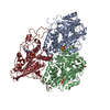
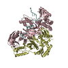
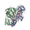
| Entry | Database: PDB / ID: 7esy | ||||||
|---|---|---|---|---|---|---|---|
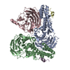
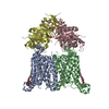
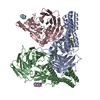
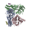
| Title | Crystal structure of the complex formed by Wolbachia cytoplasmic incompatibility factors CidA and CidBND1-ND2 from wPip | ||||||
 Components Components |
| ||||||
 Keywords Keywords |  PROTEIN BINDING / Wolbachia / deubiquitylase / antitoxin / cytoplasmic incompatibility PROTEIN BINDING / Wolbachia / deubiquitylase / antitoxin / cytoplasmic incompatibility | ||||||
| Function / homology | Ubiquitin-like protease family profile. / Ulp1 protease family, C-terminal catalytic domain / Ulp1 protease family, C-terminal catalytic domain / cysteine-type peptidase activity / Papain-like cysteine peptidase superfamily / proteolysis / CidA_IV beta/2 protein / ULP_PROTEASE domain-containing protein Function and homology information Function and homology information | ||||||
| Biological species | Wolbachia pipientis subsp. Culex pipiens | ||||||
| Method | X-RAY DIFFRACTION / SYNCHROTRON / SAD / Resolution: 2.297 Å | ||||||
 Authors Authors | Xiao, Y.J. / Wang, W. / Chen, X. / Ji, X.Y. / Yang, H.T. | ||||||
| Funding support |  China, 1items China, 1items
| ||||||
 Citation Citation | Journal: Proc.Natl.Acad.Sci.USA / Year: 2021 Title: Structural and mechanistic insights into the complexes formed by Wolbachia cytoplasmic incompatibility factors. Authors: Xiao, Y. / Chen, H. / Wang, H. / Zhang, M. / Chen, X. / Berk, J.M. / Zhang, L. / Wei, Y. / Li, W. / Cui, W. / Wang, F. / Wang, Q. / Cui, C. / Li, T. / Chen, C. / Ye, S. / Zhang, L. / Ji, X. ...Authors: Xiao, Y. / Chen, H. / Wang, H. / Zhang, M. / Chen, X. / Berk, J.M. / Zhang, L. / Wei, Y. / Li, W. / Cui, W. / Wang, F. / Wang, Q. / Cui, C. / Li, T. / Chen, C. / Ye, S. / Zhang, L. / Ji, X. / Huang, J. / Wang, W. / Wang, Z. / Hochstrasser, M. / Yang, H. | ||||||
| History |
|
- Structure visualization
| Structure viewer | Molecule: MolmilJmol/JSmol |
|---|
- Downloads & links
-Download
| PDBx/mmCIF format | 7esy.cif.gz | 463.4 KB | Display | PDBx/mmCIF format |
|---|---|---|---|---|
| PDB format | pdb7esy.ent.gz | 378.4 KB | Display | PDB format |
| PDBx/mmJSON format | 7esy.json.gz | Tree view | PDBx/mmJSON format | |
| Others |  Other downloads Other downloads |
-Validation report
| Arichive directory | https://data.pdbj.org/pub/pdb/validation_reports/es/7esyftp://data.pdbj.org/pub/pdb/validation_reports/es/7esy | HTTPS FTP |
|---|
-Related structure data


-Links
 PDBj
PDBj- Assembly
| Deposited unit | 
| ||||||||
|---|---|---|---|---|---|---|---|---|---|
| 1 |
| ||||||||
| Unit cell |
|
-Components
| #1: Protein | Mass: 57482.438 Da / Num. of mol.: 1 Source method: isolated from a genetically manipulated source Source: (gene. exp.)  Wolbachia pipientis subsp. Culex pipiens (strain wPip) (bacteria) Wolbachia pipientis subsp. Culex pipiens (strain wPip) (bacteria)Strain: wPip / Gene: WP0282 / Production host: Escherichia coli (E. coli) / References: UniProt: B3CP62 |
|---|---|
| #2: Protein | Mass: 88873.219 Da / Num. of mol.: 1 Source method: isolated from a genetically manipulated source Source: (gene. exp.) Wolbachia pipientis subsp. Culex pipiens (strain wPip) (bacteria)Strain: wPip / Gene: WP0283 / Production host: Escherichia coli (E. coli) / References: UniProt: B3CP63 |
| #3: Chemical | ChemComp-CA /   Mass: 40.078 Da / Num. of mol.: 1 / Source method: obtained synthetically / Formula: Ca Mass: 40.078 Da / Num. of mol.: 1 / Source method: obtained synthetically / Formula: Ca |
| #4: Water | ChemComp-HOH / Water Mass: 18.015 Da / Num. of mol.: 55 / Source method: isolated from a natural source / Formula: H2O Mass: 18.015 Da / Num. of mol.: 55 / Source method: isolated from a natural source / Formula: H2O |
| Has ligand of interest | N |
-Experimental details
-Experiment
| Experiment | Method: X-RAY DIFFRACTION / Number of used crystals: 1 |
|---|
- Sample preparation
| Crystal | Density Matthews: 2.29 Å3/Da / Density % sol: 46.33 % |
|---|---|
| Crystal grow | Temperature: 289 K / Method: batch mode Details: 0.2M calcium chloride dehydrate, pH 5.1, 20% w/v polyethylene glycol 3350 |
-Data collection
| Diffraction | Mean temperature: 100 K / Serial crystal experiment: N |
|---|---|
| Diffraction source | Source: SYNCHROTRON / Site: SPring-8  / Beamline: BL41XU / Wavelength: 0.97909 Å / Beamline: BL41XU / Wavelength: 0.97909 Å |
| Detector | Type: DECTRIS PILATUS3 6M / Detector: PIXEL / Date: Oct 11, 2017 |
| Radiation | Protocol: SINGLE WAVELENGTH / Monochromatic (M) / Laue (L): M / Scattering type: x-ray |
| Radiation wavelength | Wavelength: 0.97909 Å / Relative weight: 1 |
| Reflection | Resolution: 2.297→50 Å / Num. obs: 59653 / % possible obs: 96.54 % / Redundancy: 12.8 % / Biso Wilson estimate: 46.43 Å2 / Rmerge(I) obs: 0.112 / Net I/σ(I): 16.6 |
| Reflection shell | Resolution: 2.297→2.379 Å / Redundancy: 10 % / Rmerge(I) obs: 1.729 / Num. unique obs: 5741 / % possible all: 87.08 |
- Processing
| Software |
| ||||||||||||||||||||||||||||||||||||||||||||||||||||||||||||||||||
|---|---|---|---|---|---|---|---|---|---|---|---|---|---|---|---|---|---|---|---|---|---|---|---|---|---|---|---|---|---|---|---|---|---|---|---|---|---|---|---|---|---|---|---|---|---|---|---|---|---|---|---|---|---|---|---|---|---|---|---|---|---|---|---|---|---|---|---|
| Refinement | Method to determine structure: SAD / Resolution: 2.297→49.587 Å / SU ML: 0.32 / Cross valid method: THROUGHOUT / σ(F): 0 / Phase error: 28.98 / Stereochemistry target values: ML
| ||||||||||||||||||||||||||||||||||||||||||||||||||||||||||||||||||
| Solvent computation | Shrinkage radii: 0.9 Å / VDW probe radii: 1.11 Å / Solvent model: FLAT BULK SOLVENT MODEL | ||||||||||||||||||||||||||||||||||||||||||||||||||||||||||||||||||
| Displacement parameters | Biso max: 115.78 Å2 / Biso mean: 59.7603 Å2 / Biso min: 34.58 Å2 | ||||||||||||||||||||||||||||||||||||||||||||||||||||||||||||||||||
| Refinement step | Cycle: final / Resolution: 2.297→49.587 Å
| ||||||||||||||||||||||||||||||||||||||||||||||||||||||||||||||||||
| LS refinement shell | Refine-ID: X-RAY DIFFRACTION / Rfactor Rfree error: 0
| ||||||||||||||||||||||||||||||||||||||||||||||||||||||||||||||||||
| Refinement TLS params. | Method: refined / Origin x: 72.992 Å / Origin y: 39.153 Å / Origin z: 82.689 Å
| ||||||||||||||||||||||||||||||||||||||||||||||||||||||||||||||||||
| Refinement TLS group |
|