 Movie
Movie Controller

[English] 日本語
 Yorodumi
- PDB-7erp: The regulatory domain of YeiE, a sulfite sensing LysR-type transc... -
+ Open data

- Basic information
| Entry | Database: PDB / ID: 7erp | ||||||
|---|---|---|---|---|---|---|---|
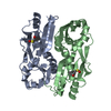
| Title | The regulatory domain of YeiE, a sulfite sensing LysR-type transcriptional regulator from Cronobacter sakazakii (sulfite-bound form) | ||||||
 Components Components | LysR family transcriptional regulator | ||||||
 Keywords Keywords |  DNA BINDING PROTEIN / LysR-type transcriptional regulator / LTTR / Cronobacter sakazakii / sulfite DNA BINDING PROTEIN / LysR-type transcriptional regulator / LTTR / Cronobacter sakazakii / sulfite | ||||||
| Function / homology |  Function and homology information Function and homology informationtranscription cis-regulatory region binding / DNA-binding transcription factor activity Similarity search - Function | ||||||
| Biological species |  Cronobacter sakazakii (bacteria) Cronobacter sakazakii (bacteria) | ||||||
| Method | X-RAY DIFFRACTION / SYNCHROTRON / MOLECULAR REPLACEMENT / Resolution: 2.03 Å | ||||||
 Authors Authors | Hong, S. / Ha, N.-C. | ||||||
| Funding support |  Korea, Republic Of, 1items Korea, Republic Of, 1items
| ||||||
 Citation Citation | Journal: Proc.Natl.Acad.Sci.USA / Year: 2022 Title: Crystal structures of YeiE from Cronobacter sakazakii and the role of sulfite tolerance in gram-negative bacteria. Authors: Hong, S. / Kim, J. / Cho, E. / Na, S. / Yoo, Y.J. / Cho, Y.H. / Ryu, S. / Ha, N.C. | ||||||
| History |
|
- Structure visualization
| Structure viewer | Molecule: MolmilJmol/JSmol |
|---|
- Downloads & links
-Download
| PDBx/mmCIF format | 7erp.cif.gz | 204.1 KB | Display | PDBx/mmCIF format |
|---|---|---|---|---|
| PDB format | pdb7erp.ent.gz | 131.8 KB | Display | PDB format |
| PDBx/mmJSON format | 7erp.json.gz | Tree view | PDBx/mmJSON format | |
| Others |  Other downloads Other downloads |
-Validation report
| Arichive directory | https://data.pdbj.org/pub/pdb/validation_reports/er/7erpftp://data.pdbj.org/pub/pdb/validation_reports/er/7erp | HTTPS FTP |
|---|
-Related structure data

-Links
 PDBj
PDBj
- Assembly
| Deposited unit | 
| ||||||||||||
|---|---|---|---|---|---|---|---|---|---|---|---|---|---|
| 1 | 
| ||||||||||||
| 2 | 
| ||||||||||||
| Unit cell |
|
-Components
| #1: Protein | Mass: 23263.971 Da / Num. of mol.: 4 Source method: isolated from a genetically manipulated source Details: sulfite / Source: (gene. exp.) Cronobacter sakazakii (bacteria) / Gene: YeiE / Production host: Escherichia coli BL21(DE3) (bacteria) / References: UniProt: H9BVC9#2: Chemical | ChemComp-SO3 / Sulfite  Mass: 80.063 Da / Num. of mol.: 4 / Source method: obtained synthetically / Formula: SO3 / Feature type: SUBJECT OF INVESTIGATION Mass: 80.063 Da / Num. of mol.: 4 / Source method: obtained synthetically / Formula: SO3 / Feature type: SUBJECT OF INVESTIGATION#3: Water | ChemComp-HOH / | Water Mass: 18.015 Da / Num. of mol.: 234 / Source method: isolated from a natural source / Formula: H2O Mass: 18.015 Da / Num. of mol.: 234 / Source method: isolated from a natural source / Formula: H2OHas ligand of interest | Y | |
|---|
-Experimental details
-Experiment
| Experiment | Method: X-RAY DIFFRACTION / Number of used crystals: 1 |
|---|
- Sample preparation
| Crystal | Density Matthews: 2.48 Å3/Da / Density % sol: 50.5 % |
|---|---|
| Crystal grow | Temperature: 287.15 K / Method: vapor diffusion, hanging drop Details: 0.1M 2-(N-morpholino)ethanesulfonic acid (MES) pH 7.0, 6% (v/v) polyethylene glycol (PEG) 20000, 2mM Tris (2-carboxyethyl) phosphine (TCEP) |
-Data collection
| Diffraction | Mean temperature: 100.15 K / Serial crystal experiment: N |
|---|---|
| Diffraction source | Source: SYNCHROTRON / Site: PAL/PLS / Beamline: 5C (4A) / Wavelength: 0.9796 Å |
| Detector | Type: DECTRIS PILATUS 6M / Detector: PIXEL / Date: Apr 30, 2016 |
| Radiation | Protocol: SINGLE WAVELENGTH / Monochromatic (M) / Laue (L): M / Scattering type: x-ray |
| Radiation wavelength | Wavelength: 0.9796 Å / Relative weight: 1 |
| Reflection | Resolution: 2.03→50 Å / Num. obs: 58388 / % possible obs: 95.8 % / Redundancy: 7.7 % / Biso Wilson estimate: 20.27 Å2 / CC star: 1 / Rmerge(I) obs: 0.049 / Rpim(I) all: 0.026 / Net I/σ(I): 18.06 |
| Reflection shell | Resolution: 2.03→2.07 Å / Redundancy: 5.4 % / Rmerge(I) obs: 0.579 / Mean I/σ(I) obs: 2.53 / Num. unique obs: 2956 / CC star: 0.817 / Rpim(I) all: 0.188 / % possible all: 98.8 |
- Processing
| Software |
| |||||||||||||||||||||||||||||||||||||||||||||||||||||||||||||||||||||||||||||||||||||||||||||||||||||||||
|---|---|---|---|---|---|---|---|---|---|---|---|---|---|---|---|---|---|---|---|---|---|---|---|---|---|---|---|---|---|---|---|---|---|---|---|---|---|---|---|---|---|---|---|---|---|---|---|---|---|---|---|---|---|---|---|---|---|---|---|---|---|---|---|---|---|---|---|---|---|---|---|---|---|---|---|---|---|---|---|---|---|---|---|---|---|---|---|---|---|---|---|---|---|---|---|---|---|---|---|---|---|---|---|---|---|---|
| Refinement | Method to determine structure: MOLECULAR REPLACEMENT Starting model: sequence based generated model Resolution: 2.03→32.28 Å / SU ML: 0.2945 / Cross valid method: FREE R-VALUE / σ(F): 1.49 / Phase error: 29.7524 Stereochemistry target values: GeoStd + Monomer Library + CDL v1.2
| |||||||||||||||||||||||||||||||||||||||||||||||||||||||||||||||||||||||||||||||||||||||||||||||||||||||||
| Solvent computation | Shrinkage radii: 0.9 Å / VDW probe radii: 1.11 Å / Solvent model: FLAT BULK SOLVENT MODEL | |||||||||||||||||||||||||||||||||||||||||||||||||||||||||||||||||||||||||||||||||||||||||||||||||||||||||
| Displacement parameters | Biso mean: 29.56 Å2 | |||||||||||||||||||||||||||||||||||||||||||||||||||||||||||||||||||||||||||||||||||||||||||||||||||||||||
| Refinement step | Cycle: LAST / Resolution: 2.03→32.28 Å
| |||||||||||||||||||||||||||||||||||||||||||||||||||||||||||||||||||||||||||||||||||||||||||||||||||||||||
| Refine LS restraints |
| |||||||||||||||||||||||||||||||||||||||||||||||||||||||||||||||||||||||||||||||||||||||||||||||||||||||||
| LS refinement shell |
|
