 Movie
Movie Controller


+ Open data

- Basic information
| Entry | Database: PDB / ID: 7epi | |||||||||
|---|---|---|---|---|---|---|---|---|---|---|
| Title | Crystal structure of E.coli CcdB mutant S60E | |||||||||
 Components Components | Toxin CcdB | |||||||||
 Keywords Keywords |  TOXIN / CcdB / Topoisomerase poison / global suppressor TOXIN / CcdB / Topoisomerase poison / global suppressor | |||||||||
| Function / homology |  Function and homology information Function and homology informationDNA topoisomerase type II (double strand cut, ATP-hydrolyzing) inhibitor activity / negative regulation of DNA-templated DNA replication / toxin-antitoxin complex / plasmid maintenance / transcription repressor complex / negative regulation of DNA-templated transcriptionSimilarity search - Function | |||||||||
| Biological species |  Escherichia coli K-12 (bacteria) Escherichia coli K-12 (bacteria) | |||||||||
| Method | X-RAY DIFFRACTION / MOLECULAR REPLACEMENT / Resolution: 1.931 Å | |||||||||
 Authors Authors | Manjunath, K. / Goyal, P. / Varadarajan, R. | |||||||||
| Funding support |  India, 2items India, 2items
| |||||||||
 Citation Citation | Journal: Plos Genet. / Year: 2022 Title: Mechanistic insights into global suppressors of protein folding defects. Authors: Chattopadhyay, G. / Bhowmick, J. / Manjunath, K. / Ahmed, S. / Goyal, P. / Varadarajan, R. | |||||||||
| History |
|
- Structure visualization
| Structure viewer | Molecule: MolmilJmol/JSmol |
|---|
- Downloads & links
-Download
| PDBx/mmCIF format | 7epi.cif.gz | 39 KB | Display | PDBx/mmCIF format |
|---|---|---|---|---|
| PDB format | pdb7epi.ent.gz | 23.4 KB | Display | PDB format |
| PDBx/mmJSON format | 7epi.json.gz | Tree view | PDBx/mmJSON format | |
| Others |  Other downloads Other downloads |
-Validation report
| Arichive directory | https://data.pdbj.org/pub/pdb/validation_reports/ep/7epiftp://data.pdbj.org/pub/pdb/validation_reports/ep/7epi | HTTPS FTP |
|---|
-Related structure data
| Related structure data | 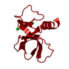 7epgC 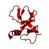 7epjC 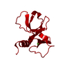 3vubS S: Starting model for refinement C: citing same article ( |
|---|---|
| Similar structure data |
-Links
 PDBj
PDBj- Assembly
| Deposited unit | 
| ||||||||||||
|---|---|---|---|---|---|---|---|---|---|---|---|---|---|
| 1 | 
| ||||||||||||
| Unit cell |
| ||||||||||||
| Components on special symmetry positions |
|
-Components
| #1: Protein | Mass: 11763.544 Da / Num. of mol.: 1 / Mutation: S60E Source method: isolated from a genetically manipulated source Source: (gene. exp.) Escherichia coli K-12 (bacteria) / Strain: K12 / Gene: ccdB, G, letB, letD, ECOK12F043 / Plasmid: pBAD24 / Production host: Escherichia coli (E. coli) / Strain (production host): Top10Gyrase / References: UniProt: P62554 | ||||
|---|---|---|---|---|---|
| #2: Chemical | Chloride  Mass: 35.453 Da / Num. of mol.: 2 / Source method: obtained synthetically / Formula: Cl Mass: 35.453 Da / Num. of mol.: 2 / Source method: obtained synthetically / Formula: Cl#3: Water | ChemComp-HOH / | Water Mass: 18.015 Da / Num. of mol.: 98 / Source method: isolated from a natural source / Formula: H2O Mass: 18.015 Da / Num. of mol.: 98 / Source method: isolated from a natural source / Formula: H2OHas ligand of interest | N | |
-Experimental details
-Experiment
| Experiment | Method: X-RAY DIFFRACTION / Number of used crystals: 1 |
|---|
- Sample preparation
| Crystal | Density Matthews: 1.9 Å3/Da / Density % sol: 35.23 % |
|---|---|
| Crystal grow | Temperature: 291 K / Method: vapor diffusion, hanging drop / pH: 5.1 / Details: 200mM CaCl2, 10% PEG 3350 |
-Data collection
| Diffraction | Mean temperature: 100 K / Serial crystal experiment: N |
|---|---|
| Diffraction source | Source: ROTATING ANODE / Type: RIGAKU MICROMAX-007 HF / Wavelength: 1.5418 Å |
| Detector | Type: MAR scanner 345 mm plate / Detector: IMAGE PLATE / Date: Dec 30, 2020 |
| Radiation | Protocol: SINGLE WAVELENGTH / Monochromatic (M) / Laue (L): M / Scattering type: x-ray |
| Radiation wavelength | Wavelength: 1.5418 Å / Relative weight: 1 |
| Reflection | Resolution: 1.93→10 Å / Num. obs: 6557 / % possible obs: 97.5 % / Redundancy: 3.4 % / Biso Wilson estimate: 8.6 Å2 / CC1/2: 0.99 / Rmerge(I) obs: 0.13 / Rpim(I) all: 0.081 / Rrim(I) all: 0.154 / Net I/σ(I): 9.6 |
| Reflection shell | Resolution: 1.93→2.03 Å / Redundancy: 3.3 % / Rmerge(I) obs: 0.424 / Mean I/σ(I) obs: 3.8 / Num. unique obs: 890 / CC1/2: 0.801 / Rpim(I) all: 0.268 / Rrim(I) all: 0.504 / % possible all: 91.6 |
- Processing
| Software |
| ||||||||||||||||||||||||||||||||||||||||||||||||||||||||||||||||||||||||||||||||||||||||||||||||||||||||||||||||||||||||||||||||||||||||||||||||||||||
|---|---|---|---|---|---|---|---|---|---|---|---|---|---|---|---|---|---|---|---|---|---|---|---|---|---|---|---|---|---|---|---|---|---|---|---|---|---|---|---|---|---|---|---|---|---|---|---|---|---|---|---|---|---|---|---|---|---|---|---|---|---|---|---|---|---|---|---|---|---|---|---|---|---|---|---|---|---|---|---|---|---|---|---|---|---|---|---|---|---|---|---|---|---|---|---|---|---|---|---|---|---|---|---|---|---|---|---|---|---|---|---|---|---|---|---|---|---|---|---|---|---|---|---|---|---|---|---|---|---|---|---|---|---|---|---|---|---|---|---|---|---|---|---|---|---|---|---|---|---|---|---|
| Refinement | Method to determine structure: MOLECULAR REPLACEMENT Starting model: 3VUB Resolution: 1.931→9.996 Å / Cor.coef. Fo:Fc: 0.929 / Cor.coef. Fo:Fc free: 0.902 / WRfactor Rfree: 0.194 / WRfactor Rwork: 0.151 / SU B: 3.531 / SU ML: 0.102 / Average fsc free: 0.9302 / Average fsc work: 0.9486 / Cross valid method: FREE R-VALUE / ESU R: 0.202 / ESU R Free: 0.162 Details: Hydrogens have been added in their riding positions
| ||||||||||||||||||||||||||||||||||||||||||||||||||||||||||||||||||||||||||||||||||||||||||||||||||||||||||||||||||||||||||||||||||||||||||||||||||||||
| Solvent computation | Ion probe radii: 0.8 Å / Shrinkage radii: 0.8 Å / VDW probe radii: 1.2 Å / Solvent model: MASK BULK SOLVENT | ||||||||||||||||||||||||||||||||||||||||||||||||||||||||||||||||||||||||||||||||||||||||||||||||||||||||||||||||||||||||||||||||||||||||||||||||||||||
| Displacement parameters | Biso mean: 10.319 Å2
| ||||||||||||||||||||||||||||||||||||||||||||||||||||||||||||||||||||||||||||||||||||||||||||||||||||||||||||||||||||||||||||||||||||||||||||||||||||||
| Refinement step | Cycle: LAST / Resolution: 1.931→9.996 Å
| ||||||||||||||||||||||||||||||||||||||||||||||||||||||||||||||||||||||||||||||||||||||||||||||||||||||||||||||||||||||||||||||||||||||||||||||||||||||
| Refine LS restraints |
| ||||||||||||||||||||||||||||||||||||||||||||||||||||||||||||||||||||||||||||||||||||||||||||||||||||||||||||||||||||||||||||||||||||||||||||||||||||||
| LS refinement shell |
|
