 Movie
Movie Controller


+ Open data

- Basic information
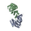
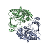
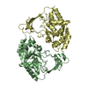
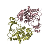
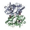
| Entry | Database: PDB / ID: 7ep0 | ||||||||||||
|---|---|---|---|---|---|---|---|---|---|---|---|---|---|
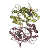
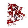
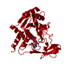
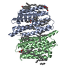
| Title | Crystal structure of ZYG11B bound to GSTE degron | ||||||||||||
 Components Components | Protein zyg-11 homolog B | ||||||||||||
 Keywords Keywords |  LIGASE / E3 ligase LIGASE / E3 ligase | ||||||||||||
| Function / homology |  Function and homology information Function and homology informationCul2-RING ubiquitin ligase complex / protein quality control for misfolded or incompletely synthesized proteins / positive regulation of proteasomal ubiquitin-dependent protein catabolic process / cytoplasmSimilarity search - Function | ||||||||||||
| Biological species |  Homo sapiens (human) Homo sapiens (human) | ||||||||||||
| Method | X-RAY DIFFRACTION / SYNCHROTRON / SAD / Resolution: 2.16 Å | ||||||||||||
 Authors Authors | Yan, X. / Li, Y. | ||||||||||||
| Funding support |  China, 3items China, 3items
| ||||||||||||
 Citation Citation | Journal: Mol.Cell / Year: 2021 Title: Molecular basis for recognition of Gly/N-degrons by CRL2 ZYG11B and CRL2 ZER1 . Authors: Yan, X. / Li, Y. / Wang, G. / Zhou, Z. / Song, G. / Feng, Q. / Zhao, Y. / Mi, W. / Ma, Z. / Dong, C. | ||||||||||||
| History |
|
- Structure visualization
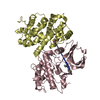
| Structure viewer | Molecule: MolmilJmol/JSmol |
|---|
- Downloads & links
-Download
| PDBx/mmCIF format | 7ep0.cif.gz | 112.2 KB | Display | PDBx/mmCIF format |
|---|---|---|---|---|
| PDB format | pdb7ep0.ent.gz | 90.6 KB | Display | PDB format |
| PDBx/mmJSON format | 7ep0.json.gz | Tree view | PDBx/mmJSON format | |
| Others |  Other downloads Other downloads |
-Validation report
| Arichive directory | https://data.pdbj.org/pub/pdb/validation_reports/ep/7ep0ftp://data.pdbj.org/pub/pdb/validation_reports/ep/7ep0 | HTTPS FTP |
|---|
-Related structure data


-Links
 PDBj
PDBj


- Assembly
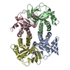
| Deposited unit | 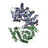
| ||||||||||||
|---|---|---|---|---|---|---|---|---|---|---|---|---|---|
| 1 |
| ||||||||||||
| Unit cell |
|
-Components
| #1: Protein | Mass: 28775.988 Da / Num. of mol.: 2 Source method: isolated from a genetically manipulated source Source: (gene. exp.) Homo sapiens (human) / Gene: ZYG11B, KIAA1730 / Production host:  Escherichia coli (E. coli) / References: UniProt: Q9C0D3 Escherichia coli (E. coli) / References: UniProt: Q9C0D3#2: Chemical | Congo red  Mass: 696.663 Da / Num. of mol.: 2 / Source method: obtained synthetically / Formula: C32H22N6Na2O6S2 Mass: 696.663 Da / Num. of mol.: 2 / Source method: obtained synthetically / Formula: C32H22N6Na2O6S2#3: Water | ChemComp-HOH / | Water Mass: 18.015 Da / Num. of mol.: 88 / Source method: isolated from a natural source / Formula: H2O Mass: 18.015 Da / Num. of mol.: 88 / Source method: isolated from a natural source / Formula: H2OHas ligand of interest | Y | |
|---|
-Experimental details
-Experiment
| Experiment | Method: X-RAY DIFFRACTION / Number of used crystals: 1 |
|---|
- Sample preparation
| Crystal | Density Matthews: 3.3 Å3/Da / Density % sol: 62.67 % |
|---|---|
| Crystal grow | Temperature: 291 K / Method: vapor diffusion, sitting drop Details: 0.1 M MES pH 6.5, 14.4% (wt/vol) Polyethylene glycol 20,000, 0.033% (wt/vol) Anthrone, 0.033% (wt/vol) Congo Red, 0.033% (wt/vol) N-(2-Acetamido)-2-aminoethanesulfonic acid, 0.002 M HEPES sodium pH 6.8 |
-Data collection
| Diffraction | Mean temperature: 80 K / Serial crystal experiment: N |
|---|---|
| Diffraction source | Source: SYNCHROTRON / Site: SSRF / Beamline: BL17U1 / Wavelength: 0.979214 Å |
| Detector | Type: MAR CCD 165 mm / Detector: CCD / Date: Nov 9, 2020 |
| Radiation | Protocol: SINGLE WAVELENGTH / Monochromatic (M) / Laue (L): M / Scattering type: x-ray |
| Radiation wavelength | Wavelength: 0.979214 Å / Relative weight: 1 |
| Reflection | Resolution: 2.16→92.98 Å / Num. obs: 39160 / % possible obs: 92.4 % / Redundancy: 15.2 % / Biso Wilson estimate: 44.79 Å2 / Rmerge(I) obs: 0.079 / Net I/σ(I): 26.7 |
| Reflection shell | Resolution: 2.16→2.27 Å / Rmerge(I) obs: 0.812 / Num. unique obs: 4143 |
- Processing
| Software |
| ||||||||||||||||||||||||
|---|---|---|---|---|---|---|---|---|---|---|---|---|---|---|---|---|---|---|---|---|---|---|---|---|---|
| Refinement | Method to determine structure: SAD / Resolution: 2.16→65.75 Å / Cross valid method: FREE R-VALUE Stereochemistry target values: GeoStd + Monomer Library + CDL v1.2
| ||||||||||||||||||||||||
| Displacement parameters | Biso mean: 50.99 Å2 | ||||||||||||||||||||||||
| Refinement step | Cycle: LAST / Resolution: 2.16→65.75 Å
| ||||||||||||||||||||||||
| Refine LS restraints |
|
