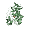
 Movie
Movie Controller


+ Open data

- Basic information
| Entry | Database: PDB / ID: 7eme | ||||||
|---|---|---|---|---|---|---|---|
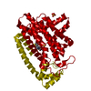
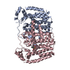
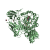
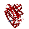
| Title | Putative Leptospira interrogans recombinant L-amino acid oxidase | ||||||
 Components Components | NAD(P)/FAD-dependent oxidoreductase | ||||||
 Keywords Keywords |  OXIDOREDUCTASE / Leptospira interrogans / L-amino acid oxidase / FAD-dependent oxidoreductase / Flavoprotein / Putative amine oxidase OXIDOREDUCTASE / Leptospira interrogans / L-amino acid oxidase / FAD-dependent oxidoreductase / Flavoprotein / Putative amine oxidase | ||||||
| Function / homology | Flavin amine oxidase / Amine oxidase / Flavin containing amine oxidoreductase / FAD/NAD(P)-binding domain superfamily / oxidoreductase activity / FLAVIN-ADENINE DINUCLEOTIDE / NAD(P)/FAD-dependent oxidoreductase Function and homology information Function and homology information | ||||||
| Biological species |  Leptospira interrogans (bacteria) Leptospira interrogans (bacteria) | ||||||
| Method | X-RAY DIFFRACTION / SYNCHROTRON / SAD / Resolution: 1.78 Å | ||||||
 Authors Authors | Vaigundan, D. / Yuvaraj, I. / Krishnaswamy, P.R. / Sekar, K. / Murthy, M.R.N. / Sunita, P. | ||||||
 Citation Citation | Journal: Curr.Sci. / Year: 2022 Title: Structural characterization of a putative recombinant L-amino acid oxidase from Leptospira interrogans Authors: Vaigundan, D. / Yuvaraj, I. / Sunita, P. / Sekar, K. / Murthy, M.R.N. / Krishnaswamy, P.R. #1: Journal: To Be PublishedTitle: Structural characterization of a putative recombinant L-amino acid oxidase from Leptospira interrogans Authors: Vaigundan, D. / Krishnaswamy, P.R. | ||||||
| History |
|
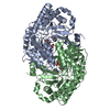
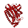
- Structure visualization
| Structure viewer | Molecule: MolmilJmol/JSmol |
|---|
- Downloads & links
-Download
| PDBx/mmCIF format | 7eme.cif.gz | 188.3 KB | Display | PDBx/mmCIF format |
|---|---|---|---|---|
| PDB format | pdb7eme.ent.gz | 145 KB | Display | PDB format |
| PDBx/mmJSON format | 7eme.json.gz | Tree view | PDBx/mmJSON format | |
| Others |  Other downloads Other downloads |
-Validation report
| Arichive directory | https://data.pdbj.org/pub/pdb/validation_reports/em/7emeftp://data.pdbj.org/pub/pdb/validation_reports/em/7eme | HTTPS FTP |
|---|
-Related structure data
| Similar structure data |
|---|


-Links
 PDBj
PDBj
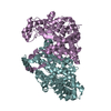
- Assembly
| Deposited unit | 
| ||||||||
|---|---|---|---|---|---|---|---|---|---|
| 1 |
| ||||||||
| Unit cell |
|
-Components
| #1: Protein | Mass: 49260.070 Da / Num. of mol.: 2 Source method: isolated from a genetically manipulated source Source: (gene. exp.) Leptospira interrogans (bacteria) / Gene: E4414_19605 / Production host: Escherichia coli BL21(DE3) (bacteria) / Variant (production host): C41 / References: UniProt: A0A4D8SV19#2: Chemical | Flavin adenine dinucleotide  Mass: 785.550 Da / Num. of mol.: 2 / Source method: obtained synthetically / Formula: C27H33N9O15P2 / Feature type: SUBJECT OF INVESTIGATION / Comment: FAD*YM Mass: 785.550 Da / Num. of mol.: 2 / Source method: obtained synthetically / Formula: C27H33N9O15P2 / Feature type: SUBJECT OF INVESTIGATION / Comment: FAD*YM#3: Water | ChemComp-HOH / | Water Mass: 18.015 Da / Num. of mol.: 737 / Source method: isolated from a natural source / Formula: H2O Mass: 18.015 Da / Num. of mol.: 737 / Source method: isolated from a natural source / Formula: H2OHas ligand of interest | Y | |
|---|
-Experimental details
-Experiment
| Experiment | Method: X-RAY DIFFRACTION / Number of used crystals: 1 |
|---|
- Sample preparation
| Crystal | Density Matthews: 1.95 Å3/Da / Density % sol: 36.99 % |
|---|---|
| Crystal grow | Temperature: 298 K / Method: microbatch Details: 0.1 M Potassium thiocyanate, 30% w/v Polyethylene glycol monomethyl ether 2000 |
-Data collection
| Diffraction | Mean temperature: 100 K / Serial crystal experiment: N |
|---|---|
| Diffraction source | Source: SYNCHROTRON / Site: ESRF  / Beamline: ID29 / Wavelength: 1.0723 Å / Beamline: ID29 / Wavelength: 1.0723 Å |
| Detector | Type: DECTRIS PILATUS3 6M / Detector: PIXEL / Date: Dec 3, 2018 |
| Radiation | Protocol: SINGLE WAVELENGTH / Monochromatic (M) / Laue (L): M / Scattering type: x-ray |
| Radiation wavelength | Wavelength: 1.0723 Å / Relative weight: 1 |
| Reflection | Resolution: 1.78→84.61 Å / Num. obs: 74369 / % possible obs: 99.1 % / Redundancy: 11.3 % / Rmerge(I) obs: 0.118 / Rpim(I) all: 0.037 / Rrim(I) all: 0.124 / Net I/σ(I): 14.3 |
| Reflection shell | Resolution: 1.78→1.81 Å / Rmerge(I) obs: 0.39 / Num. unique obs: 4197 / CC1/2: 0.978 / % possible all: 99.8 |
- Processing
| Software |
| ||||||||||||||||||||||||||||||||||||||||||||||||||||||||||||
|---|---|---|---|---|---|---|---|---|---|---|---|---|---|---|---|---|---|---|---|---|---|---|---|---|---|---|---|---|---|---|---|---|---|---|---|---|---|---|---|---|---|---|---|---|---|---|---|---|---|---|---|---|---|---|---|---|---|---|---|---|---|
| Refinement | Method to determine structure: SAD / Resolution: 1.78→70.06 Å / Cor.coef. Fo:Fc: 0.938 / Cor.coef. Fo:Fc free: 0.916 / SU B: 2.573 / SU ML: 0.082 / Cross valid method: THROUGHOUT / σ(F): 0 / ESU R: 0.133 / ESU R Free: 0.128 / Stereochemistry target values: MAXIMUM LIKELIHOOD Details: HYDROGENS HAVE BEEN ADDED IN THE RIDING POSITIONS U VALUES : REFINED INDIVIDUALLY
| ||||||||||||||||||||||||||||||||||||||||||||||||||||||||||||
| Solvent computation | Ion probe radii: 0.8 Å / Shrinkage radii: 0.8 Å / VDW probe radii: 1.2 Å / Solvent model: MASK | ||||||||||||||||||||||||||||||||||||||||||||||||||||||||||||
| Displacement parameters | Biso max: 73.44 Å2 / Biso mean: 15.409 Å2 / Biso min: 6.51 Å2
| ||||||||||||||||||||||||||||||||||||||||||||||||||||||||||||
| Refinement step | Cycle: final / Resolution: 1.78→70.06 Å
| ||||||||||||||||||||||||||||||||||||||||||||||||||||||||||||
| Refine LS restraints |
| ||||||||||||||||||||||||||||||||||||||||||||||||||||||||||||
| LS refinement shell | Resolution: 1.78→1.824 Å / Rfactor Rfree error: 0
|
