 Movie
Movie Controller


+ Open data

- Basic information
| Entry | Database: PDB / ID: 7el6 | ||||||
|---|---|---|---|---|---|---|---|
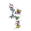
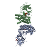
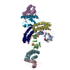
| Title | Structure of SMCR8 bound FEM1B | ||||||
 Components Components | Protein fem-1 homolog B,Guanine nucleotide exchange protein SMCR8 | ||||||
 Keywords Keywords | PEPTIDE BINDING PROTEIN / ubiquitination E3 ligase | ||||||
| Function / homology |  Function and homology information Function and homology informationregulation of ubiquitin-protein transferase activity / epithelial cell maturation involved in prostate gland development / Atg1/ULK1 kinase complex / regulation of TORC1 signaling / negative regulation of autophagosome assembly / branching involved in prostate gland morphogenesis / guanyl-nucleotide exchange factor complex /  regulation of DNA damage checkpoint / negative regulation of immune response / ubiquitin-dependent protein catabolic process via the C-end degron rule pathway ...regulation of ubiquitin-protein transferase activity / epithelial cell maturation involved in prostate gland development / Atg1/ULK1 kinase complex / regulation of TORC1 signaling / negative regulation of autophagosome assembly / branching involved in prostate gland morphogenesis / guanyl-nucleotide exchange factor complex / regulation of DNA damage checkpoint / negative regulation of immune response / ubiquitin-dependent protein catabolic process via the C-end degron rule pathway / death receptor binding / regulation of extrinsic apoptotic signaling pathway via death domain receptors / negative regulation of exocytosis / positive regulation of autophagosome maturation / Cul2-RING ubiquitin ligase complex / negative regulation of macroautophagy / protein kinase inhibitor activity / ubiquitin-like ligase-substrate adaptor activity / positive regulation of TOR signaling / GTPase activator activity / guanyl-nucleotide exchange factor activity / regulation of autophagy / cell projection / negative regulation of protein kinase activity / autophagy / positive regulation of GTPase activity / presynapse / Neddylation / postsynapse / proteasome-mediated ubiquitin-dependent protein catabolic process / protein ubiquitination / negative regulation of gene expression / apoptotic process / chromatin / protein kinase binding / nucleoplasm / metal ion binding / nucleus / cytosol / cytoplasm regulation of DNA damage checkpoint / negative regulation of immune response / ubiquitin-dependent protein catabolic process via the C-end degron rule pathway ...regulation of ubiquitin-protein transferase activity / epithelial cell maturation involved in prostate gland development / Atg1/ULK1 kinase complex / regulation of TORC1 signaling / negative regulation of autophagosome assembly / branching involved in prostate gland morphogenesis / guanyl-nucleotide exchange factor complex / regulation of DNA damage checkpoint / negative regulation of immune response / ubiquitin-dependent protein catabolic process via the C-end degron rule pathway / death receptor binding / regulation of extrinsic apoptotic signaling pathway via death domain receptors / negative regulation of exocytosis / positive regulation of autophagosome maturation / Cul2-RING ubiquitin ligase complex / negative regulation of macroautophagy / protein kinase inhibitor activity / ubiquitin-like ligase-substrate adaptor activity / positive regulation of TOR signaling / GTPase activator activity / guanyl-nucleotide exchange factor activity / regulation of autophagy / cell projection / negative regulation of protein kinase activity / autophagy / positive regulation of GTPase activity / presynapse / Neddylation / postsynapse / proteasome-mediated ubiquitin-dependent protein catabolic process / protein ubiquitination / negative regulation of gene expression / apoptotic process / chromatin / protein kinase binding / nucleoplasm / metal ion binding / nucleus / cytosol / cytoplasmSimilarity search - Function | ||||||
| Biological species |  Homo sapiens (human) Homo sapiens (human) | ||||||
| Method | X-RAY DIFFRACTION / SYNCHROTRON / MOLECULAR REPLACEMENT / Resolution: 2.802 Å | ||||||
 Authors Authors | Zhao, S. / Xu, C. | ||||||
| Funding support |  China, 1items China, 1items
| ||||||
 Citation Citation | Journal: Biochem.Biophys.Res.Commun. / Year: 2021 Title: Structural insights into SMCR8 C-degron recognition by FEM1B. Authors: Zhao, S. / Ru, W. / Chen, X. / Liao, S. / Zhu, Z. / Zhang, J. / Xu, C. | ||||||
| History |
|
- Structure visualization
| Structure viewer | Molecule: MolmilJmol/JSmol |
|---|
- Downloads & links
-Download
| PDBx/mmCIF format | 7el6.cif.gz | 269.5 KB | Display | PDBx/mmCIF format |
|---|---|---|---|---|
| PDB format | pdb7el6.ent.gz | 219.4 KB | Display | PDB format |
| PDBx/mmJSON format | 7el6.json.gz | Tree view | PDBx/mmJSON format | |
| Others |  Other downloads Other downloads |
-Validation report
| Arichive directory | https://data.pdbj.org/pub/pdb/validation_reports/el/7el6ftp://data.pdbj.org/pub/pdb/validation_reports/el/7el6 | HTTPS FTP |
|---|
-Related structure data
| Related structure data |  6lbfS S: Starting model for refinement |
|---|---|
| Similar structure data |


-Links
 PDBj
PDBj

- Assembly
| Deposited unit | 
| ||||||||
|---|---|---|---|---|---|---|---|---|---|
| 1 |
| ||||||||
| Unit cell |
|
-Components
| #1: Protein | Mass: 39301.773 Da / Num. of mol.: 2 Source method: isolated from a genetically manipulated source Details: Residues 1-337 from FEM1b, linker, residues 378-387 from SMCR8. Source: (gene. exp.) Homo sapiens (human) / Gene: FEM1B, F1AA, KIAA0396, SMCR8 / Production host:  Escherichia coli (E. coli) / References: UniProt: Q9UK73, UniProt: Q8TEV9 Escherichia coli (E. coli) / References: UniProt: Q9UK73, UniProt: Q8TEV9 |
|---|
-Experimental details
-Experiment
| Experiment | Method: X-RAY DIFFRACTION / Number of used crystals: 1 |
|---|
- Sample preparation
| Crystal | Density Matthews: 2.8 Å3/Da / Density % sol: 56.02 % |
|---|---|
| Crystal grow | Temperature: 291 K / Method: vapor diffusion, sitting drop / Details: 0.1M Tris pH7.3 4% PEG8000 |
-Data collection
| Diffraction | Mean temperature: 100 K / Serial crystal experiment: N |
|---|---|
| Diffraction source | Source: SYNCHROTRON / Site: SSRF / Beamline: BL19U1 / Wavelength: 0.97853 Å |
| Detector | Type: ADSC QUANTUM 315 / Detector: CCD / Date: Dec 6, 2020 |
| Radiation | Protocol: SINGLE WAVELENGTH / Monochromatic (M) / Laue (L): M / Scattering type: x-ray |
| Radiation wavelength | Wavelength: 0.97853 Å / Relative weight: 1 |
| Reflection | Resolution: 2.8→37.383 Å / Num. obs: 20988 / % possible obs: 100 % / Redundancy: 12.8 % / CC1/2: 1 / Net I/σ(I): 49.5 |
| Reflection shell | Resolution: 2.8→2.85 Å / Num. unique obs: 1998 / CC1/2: 0.996 |
- Processing
| Software |
| |||||||||||||||||||||||||||||||||||||||||||||||||||||||||||||||||||||||||||||||||||||||||||||||||||||||||||||||||||||||||||||
|---|---|---|---|---|---|---|---|---|---|---|---|---|---|---|---|---|---|---|---|---|---|---|---|---|---|---|---|---|---|---|---|---|---|---|---|---|---|---|---|---|---|---|---|---|---|---|---|---|---|---|---|---|---|---|---|---|---|---|---|---|---|---|---|---|---|---|---|---|---|---|---|---|---|---|---|---|---|---|---|---|---|---|---|---|---|---|---|---|---|---|---|---|---|---|---|---|---|---|---|---|---|---|---|---|---|---|---|---|---|---|---|---|---|---|---|---|---|---|---|---|---|---|---|---|---|---|
| Refinement | Method to determine structure: MOLECULAR REPLACEMENT Starting model: 6LBF Resolution: 2.802→37.383 Å / SU ML: 0.35 / Cross valid method: THROUGHOUT / σ(F): 1.34 / Phase error: 29.71 / Stereochemistry target values: ML
| |||||||||||||||||||||||||||||||||||||||||||||||||||||||||||||||||||||||||||||||||||||||||||||||||||||||||||||||||||||||||||||
| Solvent computation | Shrinkage radii: 0.9 Å / VDW probe radii: 1.11 Å / Solvent model: FLAT BULK SOLVENT MODEL | |||||||||||||||||||||||||||||||||||||||||||||||||||||||||||||||||||||||||||||||||||||||||||||||||||||||||||||||||||||||||||||
| Displacement parameters | Biso max: 173.36 Å2 / Biso mean: 63.6504 Å2 / Biso min: 28.26 Å2 | |||||||||||||||||||||||||||||||||||||||||||||||||||||||||||||||||||||||||||||||||||||||||||||||||||||||||||||||||||||||||||||
| Refinement step | Cycle: final / Resolution: 2.802→37.383 Å
| |||||||||||||||||||||||||||||||||||||||||||||||||||||||||||||||||||||||||||||||||||||||||||||||||||||||||||||||||||||||||||||
| LS refinement shell | Refine-ID: X-RAY DIFFRACTION / Rfactor Rfree error: 0
| |||||||||||||||||||||||||||||||||||||||||||||||||||||||||||||||||||||||||||||||||||||||||||||||||||||||||||||||||||||||||||||
| Refinement TLS params. | Method: refined / Refine-ID: X-RAY DIFFRACTION
| |||||||||||||||||||||||||||||||||||||||||||||||||||||||||||||||||||||||||||||||||||||||||||||||||||||||||||||||||||||||||||||
| Refinement TLS group |
|
