 Movie
Movie Controller


+ Open data

- Basic information
| Entry | Database: PDB / ID: 7ehs | ||||||
|---|---|---|---|---|---|---|---|
| Title | Levansucrase from Brenneria sp. EniD 312 | ||||||
 Components Components | Levansucrase | ||||||
 Keywords Keywords | TRANSFERASE / Levansucrase / fructosyltransferase / levan synthesis | ||||||
| Function / homology |  Function and homology information Function and homology informationlevansucrase / levansucrase activity / carbohydrate utilization / metal ion binding Similarity search - Function | ||||||
| Biological species |  Brenneria sp. EniD312 (bacteria) Brenneria sp. EniD312 (bacteria) | ||||||
| Method |  X-RAY DIFFRACTION / SYNCHROTRON / MOLECULAR REPLACEMENT / Resolution: 1.6 Å X-RAY DIFFRACTION / SYNCHROTRON / MOLECULAR REPLACEMENT / Resolution: 1.6 Å | ||||||
 Authors Authors | Xu, W. / Ni, D.W. / Hou, X.D. / Rao, Y.J. / Pijning, T. / Guskov, A. / Mu, W.M. | ||||||
| Funding support |  China, 1items China, 1items
| ||||||
 Citation Citation | Journal: J.Agric.Food Chem. / Year: 2022 Title: Crystal Structure of Levansucrase from the Gram-Negative Bacterium Brenneria Provides Insights into Its Product Size Specificity. Authors: Xu, W. / Ni, D. / Hou, X. / Pijning, T. / Guskov, A. / Rao, Y. / Mu, W. | ||||||
| History |
|
- Structure visualization
| Structure viewer | Molecule: MolmilJmol/JSmol |
|---|
- Downloads & links
-Download
| PDBx/mmCIF format | 7ehs.cif.gz | 118.4 KB | Display | PDBx/mmCIF format |
|---|---|---|---|---|
| PDB format | pdb7ehs.ent.gz | 87.8 KB | Display | PDB format |
| PDBx/mmJSON format | 7ehs.json.gz | Tree view | PDBx/mmJSON format | |
| Others |  Other downloads Other downloads |
-Validation report
| Arichive directory | https://data.pdbj.org/pub/pdb/validation_reports/eh/7ehsftp://data.pdbj.org/pub/pdb/validation_reports/eh/7ehs | HTTPS FTP |
|---|
-Related structure data
| Related structure data |  7ehrC  7ehtC  7fdzC 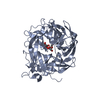 4d47S S: Starting model for refinement C: citing same article ( |
|---|---|
| Similar structure data |
-Links
 PDBj
PDBj- Assembly
| Deposited unit | 
| ||||||||
|---|---|---|---|---|---|---|---|---|---|
| 1 |
| ||||||||
| Unit cell |
| ||||||||
| Components on special symmetry positions |
|
-Components
-Protein , 1 types, 1 molecules A
| #1: Protein | Mass: 49413.855 Da / Num. of mol.: 1 / Mutation: A154S Source method: isolated from a genetically manipulated source Source: (gene. exp.) Brenneria sp. EniD312 (bacteria) / Gene: BrE312_3941 / Production host: |
|---|
-Non-polymers , 5 types, 583 molecules 


| #2: Chemical | ChemComp-PO4 /  Mass: 94.971 Da / Num. of mol.: 1 / Source method: obtained synthetically / Formula: PO4 Mass: 94.971 Da / Num. of mol.: 1 / Source method: obtained synthetically / Formula: PO4 | ||||||
|---|---|---|---|---|---|---|---|
| #3: Chemical | ChemComp-GOL /  Mass: 92.094 Da / Num. of mol.: 15 / Source method: obtained synthetically / Formula: C3H8O3 Mass: 92.094 Da / Num. of mol.: 15 / Source method: obtained synthetically / Formula: C3H8O3#4: Chemical | ChemComp-2PE / |  Mass: 414.488 Da / Num. of mol.: 1 / Source method: obtained synthetically / Formula: C18H38O10 / Comment: precipitant*YM Mass: 414.488 Da / Num. of mol.: 1 / Source method: obtained synthetically / Formula: C18H38O10 / Comment: precipitant*YM#5: Chemical |  Mass: 150.173 Da / Num. of mol.: 2 / Source method: obtained synthetically / Formula: C6H14O4 Mass: 150.173 Da / Num. of mol.: 2 / Source method: obtained synthetically / Formula: C6H14O4#6: Water | ChemComp-HOH / | Mass: 18.015 Da / Num. of mol.: 564 / Source method: isolated from a natural source / Formula: H2O |
-Details
| Has ligand of interest | N |
|---|
-Experimental details
-Experiment
| Experiment | Method: X-RAY DIFFRACTION / Number of used crystals: 1 |
|---|
- Sample preparation
| Crystal | Density Matthews: 3.25 Å3/Da / Density % sol: 62.15 % |
|---|---|
| Crystal grow | Temperature: 298 K / Method: vapor diffusion, hanging drop / Details: Ammonium sulfate, PEG 400 |
-Data collection
| Diffraction | Mean temperature: 100 K / Serial crystal experiment: N |
|---|---|
| Diffraction source | Source: SYNCHROTRON / Site: Diamond  / Beamline: I03 / Wavelength: 0.97625 Å / Beamline: I03 / Wavelength: 0.97625 Å |
| Detector | Type: DECTRIS EIGER2 XE 16M / Detector: PIXEL / Date: Jul 7, 2020 |
| Radiation | Protocol: SINGLE WAVELENGTH / Monochromatic (M) / Laue (L): M / Scattering type: x-ray |
| Radiation wavelength | Wavelength: 0.97625 Å / Relative weight: 1 |
| Reflection | Resolution: 1.34→85.15 Å / Num. obs: 80004 / % possible obs: 94.6 % / Redundancy: 39 % / Rpim(I) all: 0.025 / Net I/σ(I): 17.4 |
| Reflection shell | Resolution: 1.34→1.39 Å / Num. unique obs: 14170 / Rpim(I) all: 0.456 / % possible all: 82.3 |
- Processing
| Software |
| ||||||||||||||||||||||||||||||||||||||||||||||||||||||||||||
|---|---|---|---|---|---|---|---|---|---|---|---|---|---|---|---|---|---|---|---|---|---|---|---|---|---|---|---|---|---|---|---|---|---|---|---|---|---|---|---|---|---|---|---|---|---|---|---|---|---|---|---|---|---|---|---|---|---|---|---|---|---|
| Refinement | Method to determine structure: MOLECULAR REPLACEMENT Starting model: 4D47 Resolution: 1.6→85.14 Å / Cor.coef. Fo:Fc: 0.969 / Cor.coef. Fo:Fc free: 0.959 / WRfactor Rfree: 0.1697 / WRfactor Rwork: 0.1473 / FOM work R set: 0.8866 / SU B: 1.43 / SU ML: 0.048 / SU R Cruickshank DPI: 0.0734 / SU Rfree: 0.074 / Cross valid method: THROUGHOUT / σ(F): 0 / ESU R: 0.073 / ESU R Free: 0.074 / Stereochemistry target values: MAXIMUM LIKELIHOOD Details: HYDROGENS HAVE BEEN ADDED IN THE RIDING POSITIONS U VALUES : REFINED INDIVIDUALLY
| ||||||||||||||||||||||||||||||||||||||||||||||||||||||||||||
| Solvent computation | Ion probe radii: 0.8 Å / Shrinkage radii: 0.8 Å / VDW probe radii: 1.2 Å / Solvent model: MASK | ||||||||||||||||||||||||||||||||||||||||||||||||||||||||||||
| Displacement parameters | Biso max: 94.32 Å2 / Biso mean: 18.853 Å2 / Biso min: 8.02 Å2
| ||||||||||||||||||||||||||||||||||||||||||||||||||||||||||||
| Refinement step | Cycle: final / Resolution: 1.6→85.14 Å
| ||||||||||||||||||||||||||||||||||||||||||||||||||||||||||||
| Refine LS restraints |
| ||||||||||||||||||||||||||||||||||||||||||||||||||||||||||||
| LS refinement shell | Resolution: 1.6→1.642 Å / Rfactor Rfree error: 0 / Total num. of bins used: 20
|
