 Movie
Movie Controller

[English] 日本語
 Yorodumi
- PDB-7eey: The structure of the N-terminal doamin of the Schizosaccharomyces... -
+ Open data

- Basic information
| Entry | Database: PDB / ID: 7eey | ||||||
|---|---|---|---|---|---|---|---|
| Title | The structure of the N-terminal doamin of the Schizosaccharomyces pombe Tad2 adenosine deaminase | ||||||
 Components Components | tRNA-specific adenosine deaminase subunit tad2 | ||||||
 Keywords Keywords |  DNA BINDING PROTEIN / Tad2 / tRNA adenosine deaminase DNA BINDING PROTEIN / Tad2 / tRNA adenosine deaminase | ||||||
| Function / homology |  Function and homology information Function and homology informationtRNA-specific adenosine-34 deaminase complex / tRNA(adenine34) deaminase / tRNA wobble adenosine to inosine editing / tRNA-specific adenosine-34 deaminase activity / kinase activity / zinc ion binding / ATP binding / nucleus / cytoplasmSimilarity search - Function | ||||||
| Biological species |  Schizosaccharomyces pombe (fission yeast) Schizosaccharomyces pombe (fission yeast) | ||||||
| Method | X-RAY DIFFRACTION / SYNCHROTRON / MOLECULAR REPLACEMENT / Resolution: 2.6 Å | ||||||
 Authors Authors | Xie, W. / Liu, X. / Zhou, J. | ||||||
| Funding support |  China, 1items China, 1items
| ||||||
 Citation Citation | Journal: Comput Struct Biotechnol J / Year: 2021 Title: Functional and structural investigation of N-terminal domain of the SpTad2/3 heterodimeric tRNA deaminase. Authors: Liu, X. / Zhou, J. / Ge, R. / Xie, W. | ||||||
| History |
|
- Structure visualization
| Structure viewer | Molecule: MolmilJmol/JSmol |
|---|
- Downloads & links
-Download
| PDBx/mmCIF format | 7eey.cif.gz | 199.5 KB | Display | PDBx/mmCIF format |
|---|---|---|---|---|
| PDB format | pdb7eey.ent.gz | 127.7 KB | Display | PDB format |
| PDBx/mmJSON format | 7eey.json.gz | Tree view | PDBx/mmJSON format | |
| Others |  Other downloads Other downloads |
-Validation report
| Arichive directory | https://data.pdbj.org/pub/pdb/validation_reports/ee/7eeyftp://data.pdbj.org/pub/pdb/validation_reports/ee/7eey | HTTPS FTP |
|---|
-Related structure data
| Related structure data | 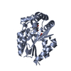 1udwS S: Starting model for refinement |
|---|---|
| Similar structure data |
-Links
 PDBj
PDBj- Assembly
| Deposited unit | 
| ||||||||||||
|---|---|---|---|---|---|---|---|---|---|---|---|---|---|
| 1 | 
| ||||||||||||
| Unit cell |
|
-Components
| #1: Protein | Mass: 22556.984 Da / Num. of mol.: 4 Source method: isolated from a genetically manipulated source Source: (gene. exp.) Schizosaccharomyces pombe (strain 972 / ATCC 24843) (yeast)Strain: 972 / ATCC 24843 / Gene: tad2, SPBC16D10.10 / Production host:  Escherichia coli (E. coli) / References: UniProt: O94642, tRNA(adenine34) deaminase Escherichia coli (E. coli) / References: UniProt: O94642, tRNA(adenine34) deaminase#2: Chemical | ChemComp-SO4 / Sulfate  Mass: 96.063 Da / Num. of mol.: 6 / Source method: obtained synthetically / Formula: SO4 / Feature type: SUBJECT OF INVESTIGATION Mass: 96.063 Da / Num. of mol.: 6 / Source method: obtained synthetically / Formula: SO4 / Feature type: SUBJECT OF INVESTIGATION#3: Water | ChemComp-HOH / | Water Mass: 18.015 Da / Num. of mol.: 167 / Source method: isolated from a natural source / Formula: H2O Mass: 18.015 Da / Num. of mol.: 167 / Source method: isolated from a natural source / Formula: H2OHas ligand of interest | Y | |
|---|
-Experimental details
-Experiment
| Experiment | Method: X-RAY DIFFRACTION / Number of used crystals: 1 |
|---|
- Sample preparation
| Crystal | Density Matthews: 2.49 Å3/Da / Density % sol: 50.56 % |
|---|---|
| Crystal grow | Temperature: 298 K / Method: vapor diffusion, hanging drop / Details: 20% PEG2000, 0.1 M NaOAc, 0.2 M (NH4)2SO4 |
-Data collection
| Diffraction | Mean temperature: 100 K / Serial crystal experiment: N |
|---|---|
| Diffraction source | Source: SYNCHROTRON / Site: SSRF / Beamline: BL19U1 / Wavelength: 0.979 Å |
| Detector | Type: DECTRIS PILATUS3 6M / Detector: PIXEL / Date: Nov 30, 2018 |
| Radiation | Protocol: SINGLE WAVELENGTH / Monochromatic (M) / Laue (L): M / Scattering type: x-ray |
| Radiation wavelength | Wavelength: 0.979 Å / Relative weight: 1 |
| Reflection | Resolution: 2.59→50 Å / Num. obs: 26761 / % possible obs: 98.7 % / Redundancy: 7.1 % / Biso Wilson estimate: 34.84 Å2 / CC1/2: 0.994 / Rmerge(I) obs: 0.07 / Net I/σ(I): 33.2 |
| Reflection shell | Resolution: 2.59→2.68 Å / Redundancy: 7.1 % / Rmerge(I) obs: 0.15 / Mean I/σ(I) obs: 15.7 / Num. unique obs: 2603 / CC1/2: 0.986 / % possible all: 97.4 |
- Processing
| Software |
| ||||||||||||||||||||||||||||||||||||||||||||||||||||||||||||||||||||||
|---|---|---|---|---|---|---|---|---|---|---|---|---|---|---|---|---|---|---|---|---|---|---|---|---|---|---|---|---|---|---|---|---|---|---|---|---|---|---|---|---|---|---|---|---|---|---|---|---|---|---|---|---|---|---|---|---|---|---|---|---|---|---|---|---|---|---|---|---|---|---|---|
| Refinement | Method to determine structure: MOLECULAR REPLACEMENT Starting model: 1UDW Resolution: 2.6→34.7 Å / SU ML: 0.2798 / Cross valid method: FREE R-VALUE / σ(F): 2.08 / Phase error: 24.7656 / Stereochemistry target values: CDL v1.2
| ||||||||||||||||||||||||||||||||||||||||||||||||||||||||||||||||||||||
| Solvent computation | Shrinkage radii: 0.9 Å / VDW probe radii: 1.11 Å / Solvent model: FLAT BULK SOLVENT MODEL | ||||||||||||||||||||||||||||||||||||||||||||||||||||||||||||||||||||||
| Displacement parameters | Biso mean: 38.53 Å2 | ||||||||||||||||||||||||||||||||||||||||||||||||||||||||||||||||||||||
| Refinement step | Cycle: LAST / Resolution: 2.6→34.7 Å
| ||||||||||||||||||||||||||||||||||||||||||||||||||||||||||||||||||||||
| Refine LS restraints |
| ||||||||||||||||||||||||||||||||||||||||||||||||||||||||||||||||||||||
| LS refinement shell |
|
