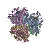
[English] 日本語
Yorodumi
- PDB-7ecr: Crystal Structure of Aspergillus terreus Glutamate Dehydrogenase ... -

+
Open data


ID or keywords:

Loading...

-
Basic information

Entry
Database: PDB / ID: 7ecr
TitleCrystal Structure of Aspergillus terreus Glutamate Dehydrogenase (AtGDH) Complexed With Succinate and ADP-ribose
ComponentsGlutamate dehydrogenase
KeywordsOXIDOREDUCTASE / Glutamate dehydrogenase / allostery / Aspergillus / enzyme mechanism
Function / homology
Function and homology information


glutamate dehydrogenase (NADP+) activity / amino acid metabolic process / nucleotide binding
Similarity search - Function
Glutamate dehydrogenase / NAD(P) binding domain of glutamate dehydrogenase / Leu/Phe/Val dehydrogenases active site / Glu / Leu / Phe / Val dehydrogenases active site. / Glutamate/phenylalanine/leucine/valine dehydrogenase / Glutamate/phenylalanine/leucine/valine dehydrogenase, dimerisation domain / Glu/Leu/Phe/Val dehydrogenase, dimerisation domain / Glutamate/Leucine/Phenylalanine/Valine dehydrogenase / Glutamate/phenylalanine/leucine/valine dehydrogenase, C-terminal / Glutamate/Leucine/Phenylalanine/Valine dehydrogenase ...Glutamate dehydrogenase / NAD(P) binding domain of glutamate dehydrogenase / Leu/Phe/Val dehydrogenases active site / Glu / Leu / Phe / Val dehydrogenases active site. / Glutamate/phenylalanine/leucine/valine dehydrogenase / Glutamate/phenylalanine/leucine/valine dehydrogenase, dimerisation domain / Glu/Leu/Phe/Val dehydrogenase, dimerisation domain / Glutamate/Leucine/Phenylalanine/Valine dehydrogenase / Glutamate/phenylalanine/leucine/valine dehydrogenase, C-terminal / Glutamate/Leucine/Phenylalanine/Valine dehydrogenase / Aminoacid dehydrogenase-like, N-terminal domain superfamily / NAD(P)-binding domain superfamily
Similarity search - Domain/homology
Chem-A2R / SUCCINIC ACID / Glutamate dehydrogenase
Similarity search - Component
Biological speciesAspergillus terreus (mold)
MethodX-RAY DIFFRACTION / MOLECULAR REPLACEMENT / Resolution: 1.73 Å
AuthorsGodsora, B.K.J. / Prakash, P. / Punekar, N.S. / Bhaumik, P.
Funding support India, 1items
OrganizationGrant numberCountry
Department of Biotechnology (DBT, India) India
CitationJournal: Proteins / Year: 2022
Title: Molecular insights into the inhibition of glutamate dehydrogenase by the dicarboxylic acid metabolites.
Authors: Godsora, B.K.J. / Prakash, P. / Punekar, N.S. / Bhaumik, P.
History
DepositionMar 13, 2021Deposition site: PDBJ / Processing site: PDBJ
Revision 1.0Dec 8, 2021Provider: repository / Type: Initial release
Revision 1.1Feb 23, 2022Group: Database references / Category: citation
Item: _citation.journal_volume / _citation.page_first ..._citation.journal_volume / _citation.page_first / _citation.page_last / _citation.year
Revision 1.2Nov 29, 2023Group: Data collection / Refinement description
Category: chem_comp_atom / chem_comp_bond / pdbx_initial_refinement_model

-
Structure visualization
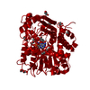
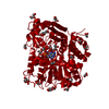
Structure viewerMolecule:
MolmilJmol/JSmol

Downloads & links

-
Assembly

Deposited unit
A: Glutamate dehydrogenase
B: Glutamate dehydrogenase
C: Glutamate dehydrogenase
hetero molecules


Theoretical massNumber of molelcules
Total (without water)151,85629
Polymers147,7423
Non-polymers4,11426
Water26,5901476
1
A: Glutamate dehydrogenase
B: Glutamate dehydrogenase
C: Glutamate dehydrogenase
hetero molecules

A: Glutamate dehydrogenase
B: Glutamate dehydrogenase
C: Glutamate dehydrogenase
hetero molecules


Theoretical massNumber of molelcules
Total (without water)303,71358
Polymers295,4856
Non-polymers8,22852
Water1086
TypeNameSymmetry operationNumber
identity operation1_555x,y,z1
crystal symmetry operation2_455-x-1,-y,z1
Buried area39190 Å2
ΔGint-8 kcal/mol
Surface area80280 Å2
MethodPISA
Unit cell
Length a, b, c (Å)119.880, 153.750, 259.950
Angle α, β, γ (deg.)90.00, 90.00, 90.00
Int Tables number23
Space group name H-MI222
Components on special symmetry positions
IDModelComponents
11C-639-

HOH

21C-1104-

HOH

-
Components

#1: Protein Glutamate dehydrogenase /


Mass: 49247.438 Da / Num. of mol.: 3
Source method: isolated from a genetically manipulated source
Details: Succinate and ADP-ribose / Source: (gene. exp.) Aspergillus terreus (mold) / Gene: gdhA, ATETN484_0007063400 / Production host: Escherichia coli (E. coli) / References: UniProt: T2D1F5
#2: Chemical
ChemComp-GOL / GLYCEROL / GLYCERIN / PROPANE-1,2,3-TRIOL / Glycerol


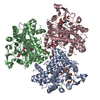
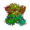
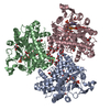
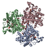
Mass: 92.094 Da / Num. of mol.: 20 / Source method: obtained synthetically / Formula: C3H8O3
#3: Chemical ChemComp-SIN / SUCCINIC ACID / Succinic acid


Mass: 118.088 Da / Num. of mol.: 3 / Source method: obtained synthetically / Formula: C4H6O4 / Feature type: SUBJECT OF INVESTIGATION
#4: Chemical ChemComp-A2R / [(2R,3R,4R,5R)-5-(6-AMINO-9H-PURIN-9-YL)-3-HYDROXY-4-(PHOSPHONOOXY)TETRAHYDROFURAN-2-YL]METHYL [(2R,3S,4R,5R)-3,4,5-TRIHYDROXYTETRAHYDROFURAN-2-YL]METHYL DIHYDROGEN DIPHOSPHATE


Mass: 639.296 Da / Num. of mol.: 3 / Source method: obtained synthetically / Formula: C15H24N5O17P3 / Feature type: SUBJECT OF INVESTIGATION
#5: Water ChemComp-HOH / water / Water


Mass: 18.015 Da / Num. of mol.: 1476 / Source method: isolated from a natural source / Formula: H2O
Has ligand of interestY

-
Experimental details

-
Experiment

ExperimentMethod: X-RAY DIFFRACTION / Number of used crystals: 1

-
Sample preparation

CrystalDensity Matthews: 4.06 Å3/Da / Density % sol: 69.68 %
Crystal growTemperature: 295 K / Method: vapor diffusion, sitting drop / pH: 7 / Details: 0.8 M Succinic acid, pH 7.0

-
Data collection

DiffractionMean temperature: 100 K / Serial crystal experiment: N
Diffraction sourceSource: ROTATING ANODE / Type: RIGAKU MICROMAX-007 HF / Wavelength: 1.5418 Å
DetectorType: RIGAKU RAXIS IV++ / Detector: IMAGE PLATE / Date: Jul 6, 2016
RadiationProtocol: SINGLE WAVELENGTH / Monochromatic (M) / Laue (L): M / Scattering type: x-ray
Radiation wavelengthWavelength: 1.5418 Å / Relative weight: 1
ReflectionResolution: 1.73→38.68 Å / Num. obs: 246054 / % possible obs: 99.2 % / Redundancy: 7.11 % / CC1/2: 0.99 / Rmerge(I) obs: 0.11 / Rrim(I) all: 0.11 / Net I/σ(I): 15.38
Reflection shellResolution: 1.73→1.83 Å / Redundancy: 5.93 % / Rmerge(I) obs: 0.91 / Mean I/σ(I) obs: 2.1 / Num. unique obs: 36141 / CC1/2: 0.72 / Rrim(I) all: 0.1 / % possible all: 94.7

-
Processing

Software
NameVersionClassification
REFMAC5.8.0189refinement
XDSdata reduction
XSCALEdata scaling
PHASERphasing
RefinementMethod to determine structure: MOLECULAR REPLACEMENT
Starting model: 5XVX
Resolution: 1.73→38.68 Å / Cor.coef. Fo:Fc: 0.975 / Cor.coef. Fo:Fc free: 0.966 / SU B: 1.649 / SU ML: 0.05 / Cross valid method: THROUGHOUT / ESU R: 0.066 / ESU R Free: 0.068 / Stereochemistry target values: MAXIMUM LIKELIHOOD / Details: HYDROGENS HAVE BEEN ADDED IN THE RIDING POSITIONS
RfactorNum. reflection% reflectionSelection details
Rfree0.167 12303 5 %RANDOM
Rwork0.145 ---
obs0.14616 233751 99.66 %-
Solvent computationIon probe radii: 0.8 Å / Shrinkage radii: 0.8 Å / VDW probe radii: 1.2 Å / Solvent model: MASK
Displacement parametersBiso mean: 19.827 Å2
Baniso -1Baniso -2Baniso -3
1--1.35 Å2-0 Å20 Å2
2--0.1 Å2-0 Å2
3---1.25 Å2
Refinement stepCycle: 1 / Resolution: 1.73→38.68 Å
ProteinNucleic acidLigandSolventTotal
Num. atoms10401 0 264 1476 12141
Refine LS restraints
Refine-IDTypeDev idealDev ideal targetNumber
X-RAY DIFFRACTIONr_bond_refined_d0.0230.01911054
X-RAY DIFFRACTIONr_bond_other_d0.0020.0210163
X-RAY DIFFRACTIONr_angle_refined_deg2.121.96914992
X-RAY DIFFRACTIONr_angle_other_deg1.101323610
X-RAY DIFFRACTIONr_dihedral_angle_1_deg5.92851439
X-RAY DIFFRACTIONr_dihedral_angle_2_deg35.53124.426479
X-RAY DIFFRACTIONr_dihedral_angle_3_deg12.162151773
X-RAY DIFFRACTIONr_dihedral_angle_4_deg15.6031563
X-RAY DIFFRACTIONr_chiral_restr0.1370.21639
X-RAY DIFFRACTIONr_gen_planes_refined0.0120.0212499
X-RAY DIFFRACTIONr_gen_planes_other0.0020.022273
X-RAY DIFFRACTIONr_nbd_refined
X-RAY DIFFRACTIONr_nbd_other
X-RAY DIFFRACTIONr_nbtor_refined
X-RAY DIFFRACTIONr_nbtor_other
X-RAY DIFFRACTIONr_xyhbond_nbd_refined
X-RAY DIFFRACTIONr_xyhbond_nbd_other
X-RAY DIFFRACTIONr_metal_ion_refined
X-RAY DIFFRACTIONr_metal_ion_other
X-RAY DIFFRACTIONr_symmetry_vdw_refined
X-RAY DIFFRACTIONr_symmetry_vdw_other
X-RAY DIFFRACTIONr_symmetry_hbond_refined
X-RAY DIFFRACTIONr_symmetry_hbond_other
X-RAY DIFFRACTIONr_symmetry_metal_ion_refined
X-RAY DIFFRACTIONr_symmetry_metal_ion_other
X-RAY DIFFRACTIONr_mcbond_it1.7561.7845590
X-RAY DIFFRACTIONr_mcbond_other1.7551.7845589
X-RAY DIFFRACTIONr_mcangle_it2.1822.6647002
X-RAY DIFFRACTIONr_mcangle_other2.1822.6647003
X-RAY DIFFRACTIONr_scbond_it3.0342.0255463
X-RAY DIFFRACTIONr_scbond_other3.0292.0255463
X-RAY DIFFRACTIONr_scangle_it
X-RAY DIFFRACTIONr_scangle_other4.2852.9367961
X-RAY DIFFRACTIONr_long_range_B_refined5.23922.82313270
X-RAY DIFFRACTIONr_long_range_B_other5.23922.82713271
X-RAY DIFFRACTIONr_rigid_bond_restr
X-RAY DIFFRACTIONr_sphericity_free
X-RAY DIFFRACTIONr_sphericity_bonded
LS refinement shellResolution: 1.733→1.778 Å / Total num. of bins used: 20
RfactorNum. reflection% reflection
Rfree0.348 868 -
Rwork0.332 16498 -
obs--95.69 %

+
About Yorodumi

-
News

-
Feb 9, 2022. New format data for meta-information of EMDB entries

New format data for meta-information of EMDB entries

  • Version 3 of the EMDB header file is now the official format.
  • The previous official version 1.9 will be removed from the archive.

Related info.:EMDB header

External links:wwPDB to switch to version 3 of the EMDB data model

-
Aug 12, 2020. Covid-19 info

Covid-19 info

URL: https://pdbj.org/emnavi/covid19.php

New page: Covid-19 featured information page in EM Navigator.

Related info.:Covid-19 info / Mar 5, 2020. Novel coronavirus structure data

+
Mar 5, 2020. Novel coronavirus structure data

Novel coronavirus structure data

Related info.:Yorodumi Speices / Aug 12, 2020. Covid-19 info

External links:COVID-19 featured content - PDBj / Molecule of the Month (242):Coronavirus Proteases

+
Jan 31, 2019. EMDB accession codes are about to change! (news from PDBe EMDB page)

EMDB accession codes are about to change! (news from PDBe EMDB page)

  • The allocation of 4 digits for EMDB accession codes will soon come to an end. Whilst these codes will remain in use, new EMDB accession codes will include an additional digit and will expand incrementally as the available range of codes is exhausted. The current 4-digit format prefixed with “EMD-” (i.e. EMD-XXXX) will advance to a 5-digit format (i.e. EMD-XXXXX), and so on. It is currently estimated that the 4-digit codes will be depleted around Spring 2019, at which point the 5-digit format will come into force.
  • The EM Navigator/Yorodumi systems omit the EMD- prefix.

Related info.:Q: What is EMD? / ID/Accession-code notation in Yorodumi/EM Navigator

External links:EMDB Accession Codes are Changing Soon! / Contact to PDBj

+
Jul 12, 2017. Major update of PDB

Major update of PDB

  • wwPDB released updated PDB data conforming to the new PDBx/mmCIF dictionary.
  • This is a major update changing the version number from 4 to 5, and with Remediation, in which all the entries are updated.
  • In this update, many items about electron microscopy experimental information are reorganized (e.g. em_software).
  • Now, EM Navigator and Yorodumi are based on the updated data.

External links:wwPDB Remediation / Enriched Model Files Conforming to OneDep Data Standards Now Available in the PDB FTP Archive

-
Yorodumi

Thousand views of thousand structures

  • Yorodumi is a browser for structure data from EMDB, PDB, SASBDB, etc.
  • This page is also the successor to EM Navigator detail page, and also detail information page/front-end page for Omokage search.
  • The word "yorodu" (or yorozu) is an old Japanese word meaning "ten thousand". "mi" (miru) is to see.

Related info.:EMDB / PDB / SASBDB / Comparison of 3 databanks / Yorodumi Search / Aug 31, 2016. New EM Navigator & Yorodumi / Yorodumi Papers / Jmol/JSmol / Function and homology information / Changes in new EM Navigator and Yorodumi

Read more