 Movie
Movie Controller

[English] 日本語
 Yorodumi
- PDB-7ebo: Crystal structure of a feruloyl esterase LP_0796 from Lactobacill... -
+ Open data

- Basic information
| Entry | Database: PDB / ID: 7ebo | ||||||
|---|---|---|---|---|---|---|---|
| Title | Crystal structure of a feruloyl esterase LP_0796 from Lactobacillus plantarum | ||||||
 Components Components | Carboxylesterase | ||||||
 Keywords Keywords | HYDROLASE / feruloyl esterase / Lactobacillus plantarum / carboxylesterase / catalytic triad | ||||||
| Function / homology | Esterase/lipase / methyl indole-3-acetate esterase activity / carboxylesterase / Serine aminopeptidase, S33 / Serine aminopeptidase, S33 / aminopeptidase activity / Alpha/Beta hydrolase fold / Carboxylesterase Function and homology information Function and homology information | ||||||
| Biological species |  Lactiplantibacillus plantarum WCFS1 (bacteria) Lactiplantibacillus plantarum WCFS1 (bacteria) | ||||||
| Method | X-RAY DIFFRACTION / SYNCHROTRON / MOLECULAR REPLACEMENT / Resolution: 2.5 Å | ||||||
 Authors Authors | Zhang, H.W. / Wang, Y.L. / Xin, F.J. | ||||||
| Funding support |  China, 1items China, 1items
| ||||||
 Citation Citation | Journal: Int.J.Biol.Macromol. / Year: 2021 Title: A reverse catalytic triad Asp containing loop shaping a wide substrate binding pocket of a feruloyl esterase from Lactobacillus plantarum. Authors: Zhang, H. / Wen, B. / Liu, Y. / Du, G. / Wei, X. / Imam, K.M.S.U. / Zhou, H. / Fan, S. / Wang, F. / Wang, Y. / Xin, F. | ||||||
| History |
|
- Structure visualization
| Structure viewer | Molecule: MolmilJmol/JSmol |
|---|
- Downloads & links
-Download
| PDBx/mmCIF format | 7ebo.cif.gz | 199.5 KB | Display | PDBx/mmCIF format |
|---|---|---|---|---|
| PDB format | pdb7ebo.ent.gz | 158.1 KB | Display | PDB format |
| PDBx/mmJSON format | 7ebo.json.gz | Tree view | PDBx/mmJSON format | |
| Others |  Other downloads Other downloads |
-Validation report
| Arichive directory | https://data.pdbj.org/pub/pdb/validation_reports/eb/7eboftp://data.pdbj.org/pub/pdb/validation_reports/eb/7ebo | HTTPS FTP |
|---|
-Related structure data
| Related structure data | 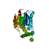 1tqhS S: Starting model for refinement |
|---|---|
| Similar structure data |
-Links
 PDBj
PDBj- Assembly
| Deposited unit | 
| ||||||||
|---|---|---|---|---|---|---|---|---|---|
| 1 |
| ||||||||
| Unit cell |
|
-Components
| #1: Protein | Mass: 27953.426 Da / Num. of mol.: 2 Source method: isolated from a genetically manipulated source Source: (gene. exp.) Lactiplantibacillus plantarum WCFS1 (bacteria)Strain: ATCC BAA-793 / NCIMB 8826 / WCFS1 / Gene: lp_0796 / Production host: Escherichia coli BL21(DE3) (bacteria) / References: UniProt: F9UM18, carboxylesterase#2: Chemical | ChemComp-SO4 / | Sulfate  Mass: 96.063 Da / Num. of mol.: 1 / Source method: obtained synthetically / Formula: SO4 / Feature type: SUBJECT OF INVESTIGATION Mass: 96.063 Da / Num. of mol.: 1 / Source method: obtained synthetically / Formula: SO4 / Feature type: SUBJECT OF INVESTIGATION#3: Water | ChemComp-HOH / | Water Mass: 18.015 Da / Num. of mol.: 96 / Source method: isolated from a natural source / Formula: H2O Mass: 18.015 Da / Num. of mol.: 96 / Source method: isolated from a natural source / Formula: H2OHas ligand of interest | Y | |
|---|
-Experimental details
-Experiment
| Experiment | Method: X-RAY DIFFRACTION / Number of used crystals: 1 |
|---|
- Sample preparation
| Crystal | Density Matthews: 2.02 Å3/Da / Density % sol: 39.12 % |
|---|---|
| Crystal grow | Temperature: 298 K / Method: vapor diffusion, hanging drop Details: 0.2 M ammonium sulfate, 0.1 M MES pH 6.5 and 25% polyethylene glycol 8000 |
-Data collection
| Diffraction | Mean temperature: 100 K / Serial crystal experiment: N | |||||||||||||||||||||||||||||||||||||||||||||||||||||||||||||||||||||||||||||||||||||||||||||||||||
|---|---|---|---|---|---|---|---|---|---|---|---|---|---|---|---|---|---|---|---|---|---|---|---|---|---|---|---|---|---|---|---|---|---|---|---|---|---|---|---|---|---|---|---|---|---|---|---|---|---|---|---|---|---|---|---|---|---|---|---|---|---|---|---|---|---|---|---|---|---|---|---|---|---|---|---|---|---|---|---|---|---|---|---|---|---|---|---|---|---|---|---|---|---|---|---|---|---|---|---|---|
| Diffraction source | Source: SYNCHROTRON / Site: SSRF / Beamline: BL17U1 / Wavelength: 0.9792 Å | |||||||||||||||||||||||||||||||||||||||||||||||||||||||||||||||||||||||||||||||||||||||||||||||||||
| Detector | Type: ADSC QUANTUM 315r / Detector: CCD / Date: Nov 7, 2019 | |||||||||||||||||||||||||||||||||||||||||||||||||||||||||||||||||||||||||||||||||||||||||||||||||||
| Radiation | Protocol: SINGLE WAVELENGTH / Monochromatic (M) / Laue (L): M / Scattering type: x-ray | |||||||||||||||||||||||||||||||||||||||||||||||||||||||||||||||||||||||||||||||||||||||||||||||||||
| Radiation wavelength | Wavelength: 0.9792 Å / Relative weight: 1 | |||||||||||||||||||||||||||||||||||||||||||||||||||||||||||||||||||||||||||||||||||||||||||||||||||
| Reflection | Resolution: 2.2→50 Å / Num. obs: 21091 / % possible obs: 96.2 % / Redundancy: 10.6 % / Biso Wilson estimate: 36.88 Å2 / Rmerge(I) obs: 0.112 / Rpim(I) all: 0.037 / Rrim(I) all: 0.118 / Χ2: 0.794 / Net I/σ(I): 9 / Num. measured all: 223710 | |||||||||||||||||||||||||||||||||||||||||||||||||||||||||||||||||||||||||||||||||||||||||||||||||||
| Reflection shell | Diffraction-ID: 1
|
- Processing
| Software |
| ||||||||||||||||||||||||||||||||||||
|---|---|---|---|---|---|---|---|---|---|---|---|---|---|---|---|---|---|---|---|---|---|---|---|---|---|---|---|---|---|---|---|---|---|---|---|---|---|
| Refinement | Method to determine structure: MOLECULAR REPLACEMENT Starting model: 1TQH Resolution: 2.5→49.18 Å / SU ML: 0.27 / Cross valid method: THROUGHOUT / σ(F): 1.35 / Phase error: 24.81 / Stereochemistry target values: ML
| ||||||||||||||||||||||||||||||||||||
| Solvent computation | Shrinkage radii: 0.9 Å / VDW probe radii: 1.11 Å / Solvent model: FLAT BULK SOLVENT MODEL | ||||||||||||||||||||||||||||||||||||
| Displacement parameters | Biso max: 127.61 Å2 / Biso mean: 40.0658 Å2 / Biso min: 16.42 Å2 | ||||||||||||||||||||||||||||||||||||
| Refinement step | Cycle: final / Resolution: 2.5→49.18 Å
| ||||||||||||||||||||||||||||||||||||
| Refine LS restraints |
| ||||||||||||||||||||||||||||||||||||
| LS refinement shell | Refine-ID: X-RAY DIFFRACTION / Rfactor Rfree error: 0
|
