 Movie
Movie Controller


+ Open data

- Basic information
| Entry | Database: PDB / ID: 7e7t | ||||||
|---|---|---|---|---|---|---|---|
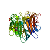
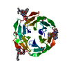
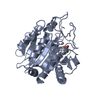
| Title | Crystal structure of RSL mutant in complex with sugar Ligand | ||||||
 Components Components | Fucose-binding lectin protein,Fucose-binding lectin protein,Fucose-binding lectin protein | ||||||
 Keywords Keywords | SUGAR BINDING PROTEIN /  Complex / Lectin / Fucose / Rhodamine Complex / Lectin / Fucose / Rhodamine | ||||||
| Function / homology | Fucose-specific lectin / Fungal fucose-specific lectin / carbohydrate binding / Chem-R2F / Fucose-binding lectin protein Function and homology information Function and homology information | ||||||
| Biological species |  Ralstonia solanacearum (bacteria) Ralstonia solanacearum (bacteria) | ||||||
| Method | X-RAY DIFFRACTION / SYNCHROTRON / MOLECULAR REPLACEMENT / Resolution: 1.98 Å | ||||||
 Authors Authors | Li, L. / Chen, G.S. | ||||||
 Citation Citation | Journal: To Be Published Title: Crystal structure of RSL mutant-R17A/R108A/R199A in complex with R2F Authors: Li, L. / Chen, G.S. | ||||||
| History |
|
- Structure visualization
| Structure viewer | Molecule: MolmilJmol/JSmol |
|---|
- Downloads & links
-Download
| PDBx/mmCIF format | 7e7t.cif.gz | 225.2 KB | Display | PDBx/mmCIF format |
|---|---|---|---|---|
| PDB format | pdb7e7t.ent.gz | 182.2 KB | Display | PDB format |
| PDBx/mmJSON format | 7e7t.json.gz | Tree view | PDBx/mmJSON format | |
| Others |  Other downloads Other downloads |
-Validation report
| Arichive directory | https://data.pdbj.org/pub/pdb/validation_reports/e7/7e7tftp://data.pdbj.org/pub/pdb/validation_reports/e7/7e7t | HTTPS FTP |
|---|
-Related structure data
| Related structure data | 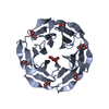 4csdS S: Starting model for refinement |
|---|---|
| Similar structure data |


-Links
 PDBj
PDBj- Assembly
| Deposited unit | 
| ||||||||||
|---|---|---|---|---|---|---|---|---|---|---|---|
| 1 | 
| ||||||||||
| 2 | 
| ||||||||||
| Unit cell |
|
-Components
| #1: Protein | Mass: 29091.568 Da / Num. of mol.: 2 / Mutation: R17A,R108A,R199A Source method: isolated from a genetically manipulated source Source: (gene. exp.) Ralstonia solanacearum (bacteria)Gene: E7Z57_08365, RSP795_21825, RSP822_19650, RUN39_v1_50103 Production host: Escherichia coli BL21(DE3) (bacteria) / References: UniProt: A0A0S4TLR1#2: Chemical | ChemComp-R2F /   Mass: 759.888 Da / Num. of mol.: 6 / Source method: obtained synthetically / Formula: C41H53N5O9 / Feature type: SUBJECT OF INVESTIGATION Mass: 759.888 Da / Num. of mol.: 6 / Source method: obtained synthetically / Formula: C41H53N5O9 / Feature type: SUBJECT OF INVESTIGATION#3: Water | ChemComp-HOH / | Water Mass: 18.015 Da / Num. of mol.: 89 / Source method: isolated from a natural source / Formula: H2O Mass: 18.015 Da / Num. of mol.: 89 / Source method: isolated from a natural source / Formula: H2OHas ligand of interest | Y | |
|---|
-Experimental details
-Experiment
| Experiment | Method: X-RAY DIFFRACTION / Number of used crystals: 1 |
|---|
- Sample preparation
| Crystal | Density Matthews: 2.37 Å3/Da / Density % sol: 48.17 % |
|---|---|
| Crystal grow | Temperature: 277 K / Method: small tubes / pH: 7.5 Details: 20 mM Tris-HCl, 100 mM of NaCl; pH 7.5, Micro centrifuge tube sequentially put with RSL solution, pure b |
-Data collection
| Diffraction | Mean temperature: 100 K / Serial crystal experiment: N | ||||||||||||||||||||||||||||||
|---|---|---|---|---|---|---|---|---|---|---|---|---|---|---|---|---|---|---|---|---|---|---|---|---|---|---|---|---|---|---|---|
| Diffraction source | Source: SYNCHROTRON / Site: SSRF  / Beamline: BL17U1 / Wavelength: 0.979183 Å / Beamline: BL17U1 / Wavelength: 0.979183 Å | ||||||||||||||||||||||||||||||
| Detector | Type: ADSC QUANTUM 315r / Detector: CCD / Date: Jan 22, 2021 | ||||||||||||||||||||||||||||||
| Radiation | Protocol: SINGLE WAVELENGTH / Monochromatic (M) / Laue (L): M / Scattering type: x-ray | ||||||||||||||||||||||||||||||
| Radiation wavelength | Wavelength: 0.979183 Å / Relative weight: 1 | ||||||||||||||||||||||||||||||
| Reflection | Resolution: 1.98→67.74 Å / Num. obs: 32990 / % possible obs: 89.6 % / Redundancy: 6.4 % / CC1/2: 0.996 / Rmerge(I) obs: 0.11 / Rpim(I) all: 0.047 / Rrim(I) all: 0.12 / Net I/σ(I): 9.2 / Num. measured all: 210690 | ||||||||||||||||||||||||||||||
| Reflection shell | Diffraction-ID: 1
|
- Processing
| Software |
| |||||||||||||||||||||||||||||||||||||||||||||||||||||||||||||||||||||||||||||||||||||||||||
|---|---|---|---|---|---|---|---|---|---|---|---|---|---|---|---|---|---|---|---|---|---|---|---|---|---|---|---|---|---|---|---|---|---|---|---|---|---|---|---|---|---|---|---|---|---|---|---|---|---|---|---|---|---|---|---|---|---|---|---|---|---|---|---|---|---|---|---|---|---|---|---|---|---|---|---|---|---|---|---|---|---|---|---|---|---|---|---|---|---|---|---|---|
| Refinement | Method to determine structure: MOLECULAR REPLACEMENT Starting model: 4CSD Resolution: 1.98→67.74 Å / SU ML: 0.31 / Cross valid method: THROUGHOUT / σ(F): 1.35 / Phase error: 36.19 / Stereochemistry target values: ML
| |||||||||||||||||||||||||||||||||||||||||||||||||||||||||||||||||||||||||||||||||||||||||||
| Solvent computation | Shrinkage radii: 0.9 Å / VDW probe radii: 1.11 Å / Solvent model: FLAT BULK SOLVENT MODEL | |||||||||||||||||||||||||||||||||||||||||||||||||||||||||||||||||||||||||||||||||||||||||||
| Displacement parameters | Biso max: 118.98 Å2 / Biso mean: 39.9042 Å2 / Biso min: 18.57 Å2 | |||||||||||||||||||||||||||||||||||||||||||||||||||||||||||||||||||||||||||||||||||||||||||
| Refinement step | Cycle: final / Resolution: 1.98→67.74 Å
| |||||||||||||||||||||||||||||||||||||||||||||||||||||||||||||||||||||||||||||||||||||||||||
| LS refinement shell | Refine-ID: X-RAY DIFFRACTION / Rfactor Rfree error: 0 / Total num. of bins used: 12
| |||||||||||||||||||||||||||||||||||||||||||||||||||||||||||||||||||||||||||||||||||||||||||
| Refinement TLS params. | Method: refined / Refine-ID: X-RAY DIFFRACTION
| |||||||||||||||||||||||||||||||||||||||||||||||||||||||||||||||||||||||||||||||||||||||||||
| Refinement TLS group |
|
