 Movie
Movie Controller

[English] 日本語
 Yorodumi
- PDB-7e50: Crystal structure of human microplasmin in complex with kazal-typ... -
+ Open data

- Basic information
| Entry | Database: PDB / ID: 7.0E+50 | ||||||
|---|---|---|---|---|---|---|---|
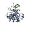
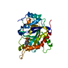
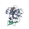
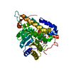
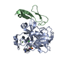
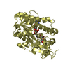
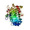
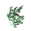
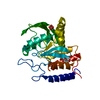
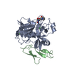
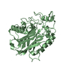
| Title | Crystal structure of human microplasmin in complex with kazal-type inhibitor AaTI | ||||||
 Components Components |
| ||||||
 Keywords Keywords |  HYDROLASE / Plasmin HYDROLASE / Plasmin | ||||||
| Function / homology |  Function and homology informationplasmin / trans-synaptic signaling by BDNF, modulating synaptic transmission / trophoblast giant cell differentiation / tissue remodeling / protein antigen binding / tissue regeneration / mononuclear cell migration / Signaling by PDGF / negative regulation of cell-cell adhesion mediated by cadherin / positive regulation of fibrinolysis ...plasmin / trans-synaptic signaling by BDNF, modulating synaptic transmission / trophoblast giant cell differentiation / tissue remodeling / protein antigen binding / tissue regeneration / mononuclear cell migration / Signaling by PDGF / negative regulation of cell-cell adhesion mediated by cadherin / positive regulation of fibrinolysis / Dissolution of Fibrin Clot / negative regulation of cell-substrate adhesion / myoblast differentiation / biological process involved in interaction with symbiont / labyrinthine layer blood vessel development / muscle cell cellular homeostasis / Activation of Matrix Metalloproteinases / apolipoprotein binding / extracellular matrix disassembly / positive regulation of blood vessel endothelial cell migration / negative regulation of fibrinolysis / fibrinolysis / serine-type peptidase activity / Degradation of the extracellular matrix / platelet alpha granule lumen / Schaffer collateral - CA1 synapse / kinase binding / Regulation of Insulin-like Growth Factor (IGF) transport and uptake by Insulin-like Growth Factor Binding Proteins (IGFBPs) / blood coagulation / Platelet degranulation / protein-folding chaperone binding / collagen-containing extracellular matrix / endopeptidase activity / protease binding / blood microparticle / protein domain specific binding / negative regulation of cell population proliferation / external side of plasma membrane / signaling receptor binding / serine-type endopeptidase activity / glutamatergic synapse / enzyme binding / cell surface / proteolysis / extracellular space / extracellular exosome / extracellular region / plasma membrane Function and homology informationplasmin / trans-synaptic signaling by BDNF, modulating synaptic transmission / trophoblast giant cell differentiation / tissue remodeling / protein antigen binding / tissue regeneration / mononuclear cell migration / Signaling by PDGF / negative regulation of cell-cell adhesion mediated by cadherin / positive regulation of fibrinolysis ...plasmin / trans-synaptic signaling by BDNF, modulating synaptic transmission / trophoblast giant cell differentiation / tissue remodeling / protein antigen binding / tissue regeneration / mononuclear cell migration / Signaling by PDGF / negative regulation of cell-cell adhesion mediated by cadherin / positive regulation of fibrinolysis / Dissolution of Fibrin Clot / negative regulation of cell-substrate adhesion / myoblast differentiation / biological process involved in interaction with symbiont / labyrinthine layer blood vessel development / muscle cell cellular homeostasis / Activation of Matrix Metalloproteinases / apolipoprotein binding / extracellular matrix disassembly / positive regulation of blood vessel endothelial cell migration / negative regulation of fibrinolysis / fibrinolysis / serine-type peptidase activity / Degradation of the extracellular matrix / platelet alpha granule lumen / Schaffer collateral - CA1 synapse / kinase binding / Regulation of Insulin-like Growth Factor (IGF) transport and uptake by Insulin-like Growth Factor Binding Proteins (IGFBPs) / blood coagulation / Platelet degranulation / protein-folding chaperone binding / collagen-containing extracellular matrix / endopeptidase activity / protease binding / blood microparticle / protein domain specific binding / negative regulation of cell population proliferation / external side of plasma membrane / signaling receptor binding / serine-type endopeptidase activity / glutamatergic synapse / enzyme binding / cell surface / proteolysis / extracellular space / extracellular exosome / extracellular region / plasma membraneSimilarity search - Function | ||||||
| Biological species |  Aedes aegypti (yellow fever mosquito) Aedes aegypti (yellow fever mosquito) Homo sapiens (human) Homo sapiens (human) | ||||||
| Method | X-RAY DIFFRACTION / SYNCHROTRON / MOLECULAR REPLACEMENT / Resolution: 1.95 Å | ||||||
 Authors Authors | Varsha, A.W. / Jobichen, C. / Mok, Y.K. | ||||||
 Citation Citation | Journal: Protein Sci. / Year: 2022 Title: Crystal structure of Aedes aegypti trypsin inhibitor in complex with mu-plasmin reveals role for scaffold stability in Kazal-type serine protease inhibitor. Authors: Walvekar, V.A. / Ramesh, K. / Jobichen, C. / Kannan, M. / Sivaraman, J. / Kini, R.M. / Mok, Y.K. | ||||||
| History |
|
- Structure visualization
| Structure viewer | Molecule: MolmilJmol/JSmol |
|---|
- Downloads & links
-Download
| PDBx/mmCIF format | 7e50.cif.gz | 138.2 KB | Display | PDBx/mmCIF format |
|---|---|---|---|---|
| PDB format | pdb7e50.ent.gz | 105.2 KB | Display | PDB format |
| PDBx/mmJSON format | 7e50.json.gz | Tree view | PDBx/mmJSON format | |
| Others |  Other downloads Other downloads |
-Validation report
| Arichive directory | https://data.pdbj.org/pub/pdb/validation_reports/e5/7e50ftp://data.pdbj.org/pub/pdb/validation_reports/e5/7e50 | HTTPS FTP |
|---|
-Related structure data
| Related structure data | 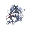 6d3xS S: Starting model for refinement |
|---|---|
| Similar structure data |


-Links
 PDBj
PDBj


- Assembly
| Deposited unit | 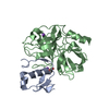
| ||||||||
|---|---|---|---|---|---|---|---|---|---|
| 1 |
| ||||||||
| Unit cell |
|
-Components
| #1: Protein | Mass: 8820.882 Da / Num. of mol.: 1 Source method: isolated from a genetically manipulated source Source: (gene. exp.) Aedes aegypti (yellow fever mosquito) / Gene: AAEL006007Production host:  Escherichia coli 'BL21-Gold(DE3)pLysS AG' (bacteria) Escherichia coli 'BL21-Gold(DE3)pLysS AG' (bacteria)References: UniProt: Q1HRB8 | ||||
|---|---|---|---|---|---|
| #2: Protein | Plasmin / MicroPlasminogen Mass: 27850.992 Da / Num. of mol.: 1 Source method: isolated from a genetically manipulated source Source: (gene. exp.) Homo sapiens (human) / Gene: PLGProduction host: Escherichia coli 'BL21-Gold(DE3)pLysS AG' (bacteria)References: UniProt: P00747, plasmin | ||||
| #3: Chemical | ChemComp-GOL / Glycerol  Mass: 92.094 Da / Num. of mol.: 1 / Source method: obtained synthetically / Formula: C3H8O3 Mass: 92.094 Da / Num. of mol.: 1 / Source method: obtained synthetically / Formula: C3H8O3 | ||||
| #4: Chemical |   Mass: 22.990 Da / Num. of mol.: 3 / Source method: obtained synthetically / Formula: Na Mass: 22.990 Da / Num. of mol.: 3 / Source method: obtained synthetically / Formula: Na#5: Water | ChemComp-HOH / | Water Mass: 18.015 Da / Num. of mol.: 138 / Source method: isolated from a natural source / Formula: H2O Mass: 18.015 Da / Num. of mol.: 138 / Source method: isolated from a natural source / Formula: H2OHas ligand of interest | N | |
-Experimental details
-Experiment
| Experiment | Method: X-RAY DIFFRACTION / Number of used crystals: 1 |
|---|
- Sample preparation
| Crystal | Density Matthews: 2.15 Å3/Da / Density % sol: 42.88 % |
|---|---|
| Crystal grow | Temperature: 296 K / Method: vapor diffusion, hanging drop Details: 0.2 M Ammonium formate, 20 % polyethylene glycol 3350 pH 6.6 |
-Data collection
| Diffraction | Mean temperature: 100 K / Serial crystal experiment: N |
|---|---|
| Diffraction source | Source: SYNCHROTRON / Site: SLS  / Beamline: X06DA / Wavelength: 0.979 Å / Beamline: X06DA / Wavelength: 0.979 Å |
| Detector | Type: DECTRIS PILATUS 2M-F / Detector: PIXEL / Date: Nov 18, 2019 |
| Radiation | Protocol: SINGLE WAVELENGTH / Monochromatic (M) / Laue (L): M / Scattering type: x-ray |
| Radiation wavelength | Wavelength: 0.979 Å / Relative weight: 1 |
| Reflection | Resolution: 1.85→44.72 Å / Num. obs: 28769 / % possible obs: 99.6 % / Redundancy: 12.4 % / Rpim(I) all: 0.057 / Net I/σ(I): 7.8 |
| Reflection shell | Resolution: 1.85→1.89 Å / Num. unique obs: 1736 / Rpim(I) all: 0.602 |
- Processing
| Software |
| |||||||||||||||||||||||||||||||||||||||||||||||||||||||||||||||||||||||||||||||||||||||||||
|---|---|---|---|---|---|---|---|---|---|---|---|---|---|---|---|---|---|---|---|---|---|---|---|---|---|---|---|---|---|---|---|---|---|---|---|---|---|---|---|---|---|---|---|---|---|---|---|---|---|---|---|---|---|---|---|---|---|---|---|---|---|---|---|---|---|---|---|---|---|---|---|---|---|---|---|---|---|---|---|---|---|---|---|---|---|---|---|---|---|---|---|---|
| Refinement | Method to determine structure: MOLECULAR REPLACEMENT Starting model: 6D3X Resolution: 1.95→19.78 Å / SU ML: 0.23 / Cross valid method: THROUGHOUT / σ(F): 1.34 / Phase error: 31.82 / Stereochemistry target values: ML
| |||||||||||||||||||||||||||||||||||||||||||||||||||||||||||||||||||||||||||||||||||||||||||
| Solvent computation | Shrinkage radii: 0.9 Å / VDW probe radii: 1.11 Å / Solvent model: FLAT BULK SOLVENT MODEL | |||||||||||||||||||||||||||||||||||||||||||||||||||||||||||||||||||||||||||||||||||||||||||
| Displacement parameters | Biso max: 104.1 Å2 / Biso mean: 49.15 Å2 / Biso min: 22.64 Å2 | |||||||||||||||||||||||||||||||||||||||||||||||||||||||||||||||||||||||||||||||||||||||||||
| Refinement step | Cycle: final / Resolution: 1.95→19.78 Å
| |||||||||||||||||||||||||||||||||||||||||||||||||||||||||||||||||||||||||||||||||||||||||||
| LS refinement shell | Refine-ID: X-RAY DIFFRACTION / Rfactor Rfree error: 0 / Total num. of bins used: 12
| |||||||||||||||||||||||||||||||||||||||||||||||||||||||||||||||||||||||||||||||||||||||||||
| Refinement TLS params. | Method: refined / Origin x: 15.4138 Å / Origin y: 23.1472 Å / Origin z: 11.1548 Å
| |||||||||||||||||||||||||||||||||||||||||||||||||||||||||||||||||||||||||||||||||||||||||||
| Refinement TLS group |
|
