 Movie
Movie Controller

[English] 日本語
 Yorodumi
- PDB-7duw: Cryo-EM structure of the multiple peptide resistance factor (MprF... -
+ Open data

- Basic information
| Entry | Database: PDB / ID: 7duw | ||||||||||||
|---|---|---|---|---|---|---|---|---|---|---|---|---|---|
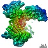
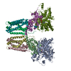
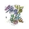
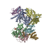
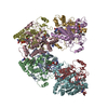
| Title | Cryo-EM structure of the multiple peptide resistance factor (MprF) loaded with two lysyl-phosphatidylglycerol molecules | ||||||||||||
 Components Components | Bifunctional lysylphosphatidylglycerol flippase/synthetase MprF | ||||||||||||
 Keywords Keywords |  MEMBRANE PROTEIN / bacteria membrane protein MEMBRANE PROTEIN / bacteria membrane protein | ||||||||||||
| Function / homology | Phosphatidylglycerol lysyltransferase, C-terminal / Phosphatidylglycerol lysyltransferase, C-terminal / Acyl-CoA N-acyltransferase / plasma membrane / Chem-EV9 / Chem-J4U / 1,2-DIPALMITOYL-PHOSPHATIDYL-GLYCEROLE / Chem-PGT / Bifunctional lysylphosphatidylglycerol flippase/synthetase MprF Function and homology information Function and homology information | ||||||||||||
| Biological species |  Rhizobium tropici (bacteria) Rhizobium tropici (bacteria) | ||||||||||||
| Method | ELECTRON MICROSCOPY / single particle reconstruction / cryo EM / Resolution: 2.96 Å | ||||||||||||
 Authors Authors | Song, D.F. / Jiao, H.Z. / Liu, Z.F. | ||||||||||||
| Funding support |  China, 3items China, 3items
| ||||||||||||
 Citation Citation | Journal: Nat Commun / Year: 2021 Title: Phospholipid translocation captured in a bifunctional membrane protein MprF. Authors: Danfeng Song / Haizhan Jiao / Zhenfeng Liu / Abstract: As a large family of membrane proteins crucial for bacterial physiology and virulence, the Multiple Peptide Resistance Factors (MprFs) utilize two separate domains to synthesize and translocate ...As a large family of membrane proteins crucial for bacterial physiology and virulence, the Multiple Peptide Resistance Factors (MprFs) utilize two separate domains to synthesize and translocate aminoacyl phospholipids to the outer leaflets of bacterial membranes. The function of MprFs enables Staphylococcus aureus and other pathogenic bacteria to acquire resistance to daptomycin and cationic antimicrobial peptides. Here we present cryo-electron microscopy structures of MprF homodimer from Rhizobium tropici (RtMprF) at two different states in complex with lysyl-phosphatidylglycerol (LysPG). RtMprF contains a membrane-embedded lipid-flippase domain with two deep cavities opening toward the inner and outer leaflets of the membrane respectively. Intriguingly, a hook-shaped LysPG molecule is trapped inside the inner cavity with its head group bent toward the outer cavity which hosts a second phospholipid-binding site. Moreover, RtMprF exhibits multiple conformational states with the synthase domain adopting distinct positions relative to the flippase domain. Our results provide a detailed framework for understanding the mechanisms of MprF-mediated modification and translocation of phospholipids. | ||||||||||||
| History |
|
- Structure visualization
| Movie |
Movie viewer |
|---|---|
| Structure viewer | Molecule: MolmilJmol/JSmol |
- Downloads & links
-Download
| PDBx/mmCIF format | 7duw.cif.gz | 294.1 KB | Display | PDBx/mmCIF format |
|---|---|---|---|---|
| PDB format | pdb7duw.ent.gz | 248.3 KB | Display | PDB format |
| PDBx/mmJSON format | 7duw.json.gz | Tree view | PDBx/mmJSON format | |
| Others |  Other downloads Other downloads |
-Validation report
| Arichive directory | https://data.pdbj.org/pub/pdb/validation_reports/du/7duwftp://data.pdbj.org/pub/pdb/validation_reports/du/7duw | HTTPS FTP |
|---|
-Related structure data
| Related structure data | 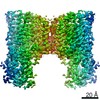 30869MC 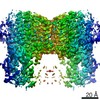 0992C 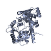 6lv0C 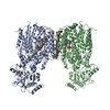 6lvfC C: citing same article ( M: map data used to model this data |
|---|---|
| Similar structure data |


-Links
 PDBj
PDBj


- Assembly
| Deposited unit | 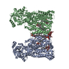
|
|---|---|
| 1 |
|
-Components
-Protein / Sugars , 2 types, 4 molecules AB
| #1: Protein | Mass: 96094.672 Da / Num. of mol.: 2 Source method: isolated from a genetically manipulated source Source: (gene. exp.) Rhizobium tropici (bacteria) / Gene: mprF, GXW80_12690 / Production host: Escherichia coli (E. coli) / References: UniProt: A0A6P1C618#2: Sugar |  Type: D-saccharide / Mass: 510.615 Da / Num. of mol.: 2 / Source method: obtained synthetically / Formula: C24H46O11 / Comment: detergent*YM Type: D-saccharide / Mass: 510.615 Da / Num. of mol.: 2 / Source method: obtained synthetically / Formula: C24H46O11 / Comment: detergent*YM |
|---|
-Non-polymers , 5 types, 12 molecules 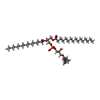

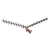
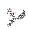

| #3: Chemical | ChemComp-EV9 / [(  Mass: 851.142 Da / Num. of mol.: 4 / Source method: obtained synthetically / Formula: C44H87N2O11P / Feature type: SUBJECT OF INVESTIGATION Mass: 851.142 Da / Num. of mol.: 4 / Source method: obtained synthetically / Formula: C44H87N2O11P / Feature type: SUBJECT OF INVESTIGATION#4: Chemical | Phosphatidylglycerol Mass: 722.970 Da / Num. of mol.: 2 / Source method: isolated from a natural source / Formula: C38H75O10P / Comment: phospholipid*YM Mass: 722.970 Da / Num. of mol.: 2 / Source method: isolated from a natural source / Formula: C38H75O10P / Comment: phospholipid*YM#5: Chemical | Phosphatidylglycerol Mass: 751.023 Da / Num. of mol.: 2 / Source method: obtained synthetically / Formula: C40H79O10P / Comment: phospholipid*YM Mass: 751.023 Da / Num. of mol.: 2 / Source method: obtained synthetically / Formula: C40H79O10P / Comment: phospholipid*YM#6: Chemical | |
|---|