 Movie
Movie Controller


+ Open data

- Basic information
| Entry | Database: PDB / ID: 7duo | ||||||
|---|---|---|---|---|---|---|---|
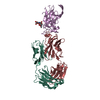
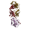
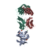
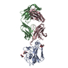
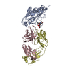
| Title | Crystal structure of daratumumab fab and CD38 complex | ||||||
 Components Components |
| ||||||
 Keywords Keywords |  ANTITUMOR PROTEIN/HYDROLASE / daratumumab / CD38 / multiple myeloma / ANTIMICROBIAL PROTEIN / ANTITUMOR PROTEIN-HYDROLASE complex ANTITUMOR PROTEIN/HYDROLASE / daratumumab / CD38 / multiple myeloma / ANTIMICROBIAL PROTEIN / ANTITUMOR PROTEIN-HYDROLASE complex | ||||||
| Function / homology |  Function and homology information Function and homology information2'-phospho-ADP-ribosyl cyclase/2'-phospho-cyclic-ADP-ribose transferase / phosphorus-oxygen lyase activity / artery smooth muscle contraction / Nicotinate metabolism / NAD+ nucleosidase activity / ADP-ribosyl cyclase/cyclic ADP-ribose hydrolase / NAD metabolic process / NAD+ nucleotidase, cyclic ADP-ribose generating / NADP+ nucleosidase activity / negative regulation of bone resorption ...2'-phospho-ADP-ribosyl cyclase/2'-phospho-cyclic-ADP-ribose transferase / phosphorus-oxygen lyase activity / artery smooth muscle contraction / Nicotinate metabolism / NAD+ nucleosidase activity / ADP-ribosyl cyclase/cyclic ADP-ribose hydrolase / NAD metabolic process / NAD+ nucleotidase, cyclic ADP-ribose generating / NADP+ nucleosidase activity / negative regulation of bone resorption / response to hydroperoxide / long-term synaptic depression / Hydrolases; Glycosylases; Hydrolysing N-glycosyl compounds / B cell proliferation / response to retinoic acid / positive regulation of B cell proliferation / positive regulation of vasoconstriction / response to interleukin-1 / response to progesterone / female pregnancy / apoptotic signaling pathway / B cell receptor signaling pathway / positive regulation of insulin secretion / response to estradiol / negative regulation of neuron projection development / positive regulation of cytosolic calcium ion concentration / transferase activity / positive regulation of cell growth / basolateral plasma membrane / response to hypoxia / response to xenobiotic stimulus / negative regulation of DNA-templated transcription / negative regulation of apoptotic process / positive regulation of DNA-templated transcription / cell surface / signal transduction / extracellular exosome / membrane / identical protein binding / plasma membraneSimilarity search - Function | ||||||
| Biological species |  Homo sapiens (human) Homo sapiens (human) | ||||||
| Method | X-RAY DIFFRACTION / SYNCHROTRON / MOLECULAR REPLACEMENT / Resolution: 2.81 Å | ||||||
 Authors Authors | Yu, X.J. / Wang, L. / Yu, C.F. | ||||||
 Citation Citation | Journal: To Be Published Title: Crystal structure of daratumumab fab and CD38 complex Authors: Yu, X.J. / Wang, L. / Yu, C.F. | ||||||
| History |
|
- Structure visualization
| Structure viewer | Molecule: MolmilJmol/JSmol |
|---|
- Downloads & links
-Download
| PDBx/mmCIF format | 7duo.cif.gz | 259.9 KB | Display | PDBx/mmCIF format |
|---|---|---|---|---|
| PDB format | pdb7duo.ent.gz | 209.1 KB | Display | PDB format |
| PDBx/mmJSON format | 7duo.json.gz | Tree view | PDBx/mmJSON format | |
| Others |  Other downloads Other downloads |
-Validation report
| Arichive directory | https://data.pdbj.org/pub/pdb/validation_reports/du/7duoftp://data.pdbj.org/pub/pdb/validation_reports/du/7duo | HTTPS FTP |
|---|
-Related structure data
| Related structure data | 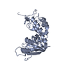 1yh3S S: Starting model for refinement |
|---|---|
| Similar structure data |


-Links
 PDBj
PDBj


- Assembly
| Deposited unit | 
| ||||||||
|---|---|---|---|---|---|---|---|---|---|
| 1 |
| ||||||||
| Unit cell |
|
-Components
| #1: Antibody | Fragment antigen-binding Mass: 23595.488 Da / Num. of mol.: 1 Source method: isolated from a genetically manipulated source Source: (gene. exp.) Homo sapiens (human) / Production host:   Cricetulus griseus (Chinese hamster) Cricetulus griseus (Chinese hamster) |
|---|---|
| #2: Protein | Mass: 26650.408 Da / Num. of mol.: 1 Source method: isolated from a genetically manipulated source Source: (gene. exp.) Homo sapiens (human) / Gene: CD38Production host: Mammalian expression vector BsrGI-MCS-pcDNA3.1 (others) References: UniProt: P28907, ADP-ribosyl cyclase/cyclic ADP-ribose hydrolase, 2'-phospho-ADP-ribosyl cyclase/2'-phospho-cyclic-ADP-ribose transferase |
| #3: Antibody | Fragment antigen-binding Mass: 23302.799 Da / Num. of mol.: 1 Source method: isolated from a genetically manipulated source Source: (gene. exp.) Homo sapiens (human) / Production host: Cricetulus griseus (Chinese hamster) |
-Experimental details
-Experiment
| Experiment | Method: X-RAY DIFFRACTION / Number of used crystals: 1 |
|---|
- Sample preparation
| Crystal | Density Matthews: 2.79 Å3/Da / Density % sol: 55.96 % |
|---|---|
| Crystal grow | Temperature: 291 K / Method: evaporation, recrystallization Details: 0.2M Sodium phosphate monobasic monohydrate, 20% w/v Polyethylene glycol 3350 |
-Data collection
| Diffraction | Mean temperature: 100 K / Serial crystal experiment: N |
|---|---|
| Diffraction source | Source: SYNCHROTRON / Site: SSRF  / Beamline: BL17U / Wavelength: 0.9798 Å / Beamline: BL17U / Wavelength: 0.9798 Å |
| Detector | Type: DECTRIS EIGER2 X 16M / Detector: PIXEL / Date: Sep 16, 2020 |
| Radiation | Protocol: SINGLE WAVELENGTH / Monochromatic (M) / Laue (L): M / Scattering type: x-ray |
| Radiation wavelength | Wavelength: 0.9798 Å / Relative weight: 1 |
| Reflection | Resolution: 2.81→50 Å / Num. obs: 19416 / % possible obs: 100 % / Redundancy: 6.4 % / CC1/2: 0.969 / Rmerge(I) obs: 0.183 / Net I/σ(I): 12.18 |
| Reflection shell | Resolution: 2.81→2.9 Å / Rmerge(I) obs: 0.183 / Num. unique obs: 19416 / CC1/2: 0.969 |
- Processing
| Software |
| ||||||||||||||||||||||||||||||||||||||||||||||||||||||||||||||||||||||||||||||||||||||||||
|---|---|---|---|---|---|---|---|---|---|---|---|---|---|---|---|---|---|---|---|---|---|---|---|---|---|---|---|---|---|---|---|---|---|---|---|---|---|---|---|---|---|---|---|---|---|---|---|---|---|---|---|---|---|---|---|---|---|---|---|---|---|---|---|---|---|---|---|---|---|---|---|---|---|---|---|---|---|---|---|---|---|---|---|---|---|---|---|---|---|---|---|
| Refinement | Method to determine structure: MOLECULAR REPLACEMENT Starting model: 1yh3 Resolution: 2.81→46.055 Å / SU ML: 0.34 / Cross valid method: THROUGHOUT / σ(F): 1.37 / Phase error: 26.5 / Stereochemistry target values: ML
| ||||||||||||||||||||||||||||||||||||||||||||||||||||||||||||||||||||||||||||||||||||||||||
| Solvent computation | Shrinkage radii: 0.9 Å / VDW probe radii: 1.11 Å / Solvent model: FLAT BULK SOLVENT MODEL | ||||||||||||||||||||||||||||||||||||||||||||||||||||||||||||||||||||||||||||||||||||||||||
| Displacement parameters | Biso max: 124.93 Å2 / Biso mean: 36.1454 Å2 / Biso min: 7.85 Å2 | ||||||||||||||||||||||||||||||||||||||||||||||||||||||||||||||||||||||||||||||||||||||||||
| Refinement step | Cycle: final / Resolution: 2.81→46.055 Å
| ||||||||||||||||||||||||||||||||||||||||||||||||||||||||||||||||||||||||||||||||||||||||||
| LS refinement shell | Refine-ID: X-RAY DIFFRACTION / Rfactor Rfree error: 0
| ||||||||||||||||||||||||||||||||||||||||||||||||||||||||||||||||||||||||||||||||||||||||||
| Refinement TLS params. | Method: refined / Origin x: 39.1934 Å / Origin y: 30.0133 Å / Origin z: 21.1732 Å
| ||||||||||||||||||||||||||||||||||||||||||||||||||||||||||||||||||||||||||||||||||||||||||
| Refinement TLS group |
|
